 Movie
Movie Controller

[English] 日本語
 Yorodumi
- PDB-5jnr: Crystal structure of human low molecular weight protein tyrosine ... -
+ Open data

- Basic information
| Entry | Database: PDB / ID: 5jnr | ||||||
|---|---|---|---|---|---|---|---|
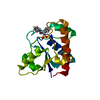
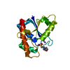
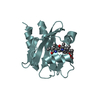
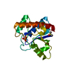
| Title | Crystal structure of human low molecular weight protein tyrosine phosphatase (LMPTP) type A | ||||||
 Components Components | Low molecular weight phosphotyrosine protein phosphatase | ||||||
 Keywords Keywords |  HYDROLASE / protein tyrosine phosphatase / LMW-PTP / LMPTP HYDROLASE / protein tyrosine phosphatase / LMW-PTP / LMPTP | ||||||
| Function / homology |  Function and homology informationacid phosphatase / acid phosphatase activity / non-membrane spanning protein tyrosine phosphatase activity / protein-tyrosine-phosphatase / protein tyrosine phosphatase activity / sarcolemma / cytoplasmic side of plasma membrane / extracellular exosome / cytosol / cytoplasm Function and homology informationacid phosphatase / acid phosphatase activity / non-membrane spanning protein tyrosine phosphatase activity / protein-tyrosine-phosphatase / protein tyrosine phosphatase activity / sarcolemma / cytoplasmic side of plasma membrane / extracellular exosome / cytosol / cytoplasmSimilarity search - Function | ||||||
| Biological species |  Homo sapiens (human) Homo sapiens (human) | ||||||
| Method | X-RAY DIFFRACTION / SYNCHROTRON / MOLECULAR REPLACEMENT / Resolution: 2 Å | ||||||
 Authors Authors | Stanford, S.M. / Aleshin, A.E. / Liddington, R.C. / Bankston, L. / Cadwell, G. / Bottini, N. | ||||||
| Funding support |  United States, 1items United States, 1items
| ||||||
 Citation Citation | Journal: Nat. Chem. Biol. / Year: 2017 Title: Diabetes reversal by inhibition of the low-molecular-weight tyrosine phosphatase. Authors: Stanford, S.M. / Aleshin, A.E. / Zhang, V. / Ardecky, R.J. / Hedrick, M.P. / Zou, J. / Ganji, S.R. / Bliss, M.R. / Yamamoto, F. / Bobkov, A.A. / Kiselar, J. / Liu, Y. / Cadwell, G.W. / ...Authors: Stanford, S.M. / Aleshin, A.E. / Zhang, V. / Ardecky, R.J. / Hedrick, M.P. / Zou, J. / Ganji, S.R. / Bliss, M.R. / Yamamoto, F. / Bobkov, A.A. / Kiselar, J. / Liu, Y. / Cadwell, G.W. / Khare, S. / Yu, J. / Barquilla, A. / Chung, T.D.Y. / Mustelin, T. / Schenk, S. / Bankston, L.A. / Liddington, R.C. / Pinkerton, A.B. / Bottini, N. | ||||||
| History |
|
- Structure visualization
| Structure viewer | Molecule: MolmilJmol/JSmol |
|---|
- Downloads & links
-Download
| PDBx/mmCIF format | 5jnr.cif.gz | 51.8 KB | Display | PDBx/mmCIF format |
|---|---|---|---|---|
| PDB format | pdb5jnr.ent.gz | 35.2 KB | Display | PDB format |
| PDBx/mmJSON format | 5jnr.json.gz | Tree view | PDBx/mmJSON format | |
| Others |  Other downloads Other downloads |
-Validation report
| Arichive directory | https://data.pdbj.org/pub/pdb/validation_reports/jn/5jnrftp://data.pdbj.org/pub/pdb/validation_reports/jn/5jnr | HTTPS FTP |
|---|
-Related structure data
| Related structure data |  5jnsC 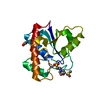 5jntC  5jnuC 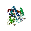 5jnvC  5jnwC  3n8iS C: citing same article ( S: Starting model for refinement |
|---|---|
| Similar structure data |


-Links
 PDBj
PDBj

- Assembly
| Deposited unit | 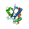
| ||||||||
|---|---|---|---|---|---|---|---|---|---|
| 1 |
| ||||||||
| Unit cell |
|
-Components
| #1: Protein | Mass: 18209.635 Da / Num. of mol.: 1 Source method: isolated from a genetically manipulated source Source: (gene. exp.) Homo sapiens (human) / Gene: ACP1 / Plasmid: pGEX-4T / Production host:  Escherichia coli (E. coli) / Strain (production host): BL21(DE3) / References: UniProt: P24666, protein-tyrosine-phosphatase Escherichia coli (E. coli) / Strain (production host): BL21(DE3) / References: UniProt: P24666, protein-tyrosine-phosphatase |
|---|---|
| #2: Water | ChemComp-HOH / Water Mass: 18.015 Da / Num. of mol.: 157 / Source method: isolated from a natural source / Formula: H2O Mass: 18.015 Da / Num. of mol.: 157 / Source method: isolated from a natural source / Formula: H2O |
-Experimental details
-Experiment
| Experiment | Method: X-RAY DIFFRACTION / Number of used crystals: 1 |
|---|
- Sample preparation
| Crystal | Density Matthews: 2.4 Å3/Da / Density % sol: 48.69 % / Mosaicity: 0.48 ° |
|---|---|
| Crystal grow | Temperature: 293 K / Method: evaporation / pH: 5.5 / Details: 7% PEG 10K, Bis Tris, Ammonium Acetate |
-Data collection
| Diffraction | Mean temperature: 100 K |
|---|---|
| Diffraction source | Source: SYNCHROTRON / Site: SSRL / Beamline: BL9-2 / Wavelength: 1.5418 Å |
| Detector | Type: ADSC QUANTUM 315r / Detector: CCD / Date: Sep 14, 2014 |
| Radiation | Monochromator: mirrows / Protocol: SINGLE WAVELENGTH / Monochromatic (M) / Laue (L): M / Scattering type: x-ray |
| Radiation wavelength | Wavelength: 1.5418 Å / Relative weight: 1 |
| Reflection | Resolution: 2→36.51 Å / Num. obs: 12105 / % possible obs: 96.9 % / Redundancy: 3.8 % / CC1/2: 0.997 / Rmerge(I) obs: 0.064 / Net I/σ(I): 34 |
| Reflection shell | Resolution: 2→2.05 Å / Redundancy: 3.4 % / Rmerge(I) obs: 0.238 / % possible all: 91.1 |
- Processing
| Software |
| |||||||||||||||||||||||||||||||||||||||||||||||||||||||||||||||||||||||||||
|---|---|---|---|---|---|---|---|---|---|---|---|---|---|---|---|---|---|---|---|---|---|---|---|---|---|---|---|---|---|---|---|---|---|---|---|---|---|---|---|---|---|---|---|---|---|---|---|---|---|---|---|---|---|---|---|---|---|---|---|---|---|---|---|---|---|---|---|---|---|---|---|---|---|---|---|---|
| Refinement | Method to determine structure: MOLECULAR REPLACEMENT Starting model: 3N8I Resolution: 2→36.51 Å / Cor.coef. Fo:Fc: 0.957 / Cor.coef. Fo:Fc free: 0.927 / SU B: 3.807 / SU ML: 0.105 / Cross valid method: THROUGHOUT / σ(F): 0 / ESU R: 0.176 / ESU R Free: 0.159 Details: HYDROGENS HAVE BEEN ADDED IN THE RIDING POSITIONS U VALUES : REFINED INDIVIDUALLY
| |||||||||||||||||||||||||||||||||||||||||||||||||||||||||||||||||||||||||||
| Solvent computation | Ion probe radii: 0.8 Å / Shrinkage radii: 0.8 Å / VDW probe radii: 1.2 Å | |||||||||||||||||||||||||||||||||||||||||||||||||||||||||||||||||||||||||||
| Displacement parameters | Biso max: 74.95 Å2 / Biso mean: 25.942 Å2 / Biso min: 7.94 Å2
| |||||||||||||||||||||||||||||||||||||||||||||||||||||||||||||||||||||||||||
| Refinement step | Cycle: final / Resolution: 2→36.51 Å
| |||||||||||||||||||||||||||||||||||||||||||||||||||||||||||||||||||||||||||
| Refine LS restraints |
| |||||||||||||||||||||||||||||||||||||||||||||||||||||||||||||||||||||||||||
| LS refinement shell | Resolution: 1.995→2.047 Å / Total num. of bins used: 20
|
