 Movie
Movie Controller


+ Open data

- Basic information
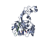
| Entry | Database: PDB / ID: 5jjy | ||||||
|---|---|---|---|---|---|---|---|
| Title | Crystal structure of SETD2 bound to histone H3.3 K36M peptide | ||||||
 Components Components |
| ||||||
 Keywords Keywords |  TRANSFERASE / SET domain TRANSFERASE / SET domain | ||||||
| Function / homology |  Function and homology information Function and homology informationmesoderm morphogenesis / coronary vasculature morphogenesis / morphogenesis of a branching structure / peptidyl-lysine trimethylation / cell migration involved in vasculogenesis / microtubule cytoskeleton organization involved in mitosis / [histone H3]-lysine36 N-trimethyltransferase / histone H3K36 trimethyltransferase activity / embryonic placenta morphogenesis / regulation of mRNA export from nucleus ...mesoderm morphogenesis / coronary vasculature morphogenesis / morphogenesis of a branching structure / peptidyl-lysine trimethylation / cell migration involved in vasculogenesis / microtubule cytoskeleton organization involved in mitosis / [histone H3]-lysine36 N-trimethyltransferase / histone H3K36 trimethyltransferase activity / embryonic placenta morphogenesis / regulation of mRNA export from nucleus / pericardium development / nucleosome organization / stem cell development / histone H3K36 methyltransferase activity / protein-lysine N-methyltransferase activity / response to type I interferon / positive regulation of ossification / embryonic cranial skeleton morphogenesis / response to alkaloid / regulation of protein localization to chromatin / response to metal ion / histone H3 methyltransferase activity / regulation of double-strand break repair via homologous recombination / endodermal cell differentiation / positive regulation of interferon-alpha production / alpha-tubulin binding / nucleosomal DNA binding / mismatch repair / RNA polymerase II core promoter sequence-specific DNA binding / Replacement of protamines by nucleosomes in the male pronucleus / positive regulation of autophagy / forebrain development / Inhibition of DNA recombination at telomere / telomere organization / RNA Polymerase I Promoter Opening / Assembly of the ORC complex at the origin of replication / Transferases; Transferring one-carbon groups; Methyltransferases / DNA methylation / Condensation of Prophase Chromosomes / ERCC6 (CSB) and EHMT2 (G9a) positively regulate rRNA expression / SIRT1 negatively regulates rRNA expression / Chromatin modifications during the maternal to zygotic transition (MZT) / regulation of cytokinesis / PRC2 methylates histones and DNA / Defective pyroptosis / neural tube closure / transcription elongation by RNA polymerase II / stem cell differentiation / RNA Polymerase I Promoter Escape / Transcriptional regulation by small RNAs / Formation of the beta-catenin:TCF transactivating complex / RUNX1 regulates genes involved in megakaryocyte differentiation and platelet function / Activated PKN1 stimulates transcription of AR (androgen receptor) regulated genes KLK2 and KLK3 / NoRC negatively regulates rRNA expression / B-WICH complex positively regulates rRNA expression / response to organic cyclic compound / PKMTs methylate histone lysines / Meiotic recombination / Pre-NOTCH Transcription and Translation / Activation of anterior HOX genes in hindbrain development during early embryogenesis / Transcriptional regulation of granulopoiesis / structural constituent of chromatin / nucleosome / nucleosome assembly / RUNX1 regulates transcription of genes involved in differentiation of HSCs / chromosome / Factors involved in megakaryocyte development and platelet production / Senescence-Associated Secretory Phenotype (SASP) / positive regulation of cell growth / regulation of gene expression / angiogenesis / Oxidative Stress Induced Senescence / defense response to virus / Estrogen-dependent gene expression / chromosome, telomeric region / RNA polymerase II cis-regulatory region sequence-specific DNA binding / protein heterodimerization activity / Amyloid fiber formation / regulation of DNA-templated transcription / protein-containing complex / extracellular exosome / extracellular region / nucleoplasm / metal ion binding / nucleus / cytoplasmSimilarity search - Function | ||||||
| Biological species |  Homo sapiens (human) Homo sapiens (human) | ||||||
| Method | X-RAY DIFFRACTION / SYNCHROTRON / MOLECULAR REPLACEMENT / molecular replacement / Resolution: 2.053 Å | ||||||
 Authors Authors | Yang, S. / Zheng, X. / Li, H. | ||||||
 Citation Citation | Journal: Genes Dev. / Year: 2016 Title: Molecular basis for oncohistone H3 recognition by SETD2 methyltransferase Authors: Yang, S. / Zheng, X. / Lu, C. / Li, G.M. / Allis, C.D. / Li, H. | ||||||
| History |
|
- Structure visualization
| Structure viewer | Molecule: MolmilJmol/JSmol |
|---|
- Downloads & links
-Download
| PDBx/mmCIF format | 5jjy.cif.gz | 76.9 KB | Display | PDBx/mmCIF format |
|---|---|---|---|---|
| PDB format | pdb5jjy.ent.gz | 53.2 KB | Display | PDB format |
| PDBx/mmJSON format | 5jjy.json.gz | Tree view | PDBx/mmJSON format | |
| Others |  Other downloads Other downloads |
-Validation report
| Arichive directory | https://data.pdbj.org/pub/pdb/validation_reports/jj/5jjyftp://data.pdbj.org/pub/pdb/validation_reports/jj/5jjy | HTTPS FTP |
|---|
-Related structure data
| Related structure data | 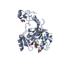 5jlbC  5jleC  4h12S C: citing same article ( S: Starting model for refinement |
|---|---|
| Similar structure data |


-Links
 PDBj
PDBj


- Assembly
| Deposited unit | 
| ||||||||
|---|---|---|---|---|---|---|---|---|---|
| 1 |
| ||||||||
| Unit cell |
|
-Components
-Protein / Protein/peptide , 2 types, 2 molecules AB
| #1: Protein | Mass: 32042.568 Da / Num. of mol.: 1 / Fragment: catalytic domain, UNP residues 1434-1711 Source method: isolated from a genetically manipulated source Source: (gene. exp.) Homo sapiens (human) / Gene: SETD2 / Plasmid: pET28b / Production host:  Escherichia coli BL21(DE3) (bacteria) / Strain (production host): BL21(DE3) Escherichia coli BL21(DE3) (bacteria) / Strain (production host): BL21(DE3)References: UniProt: Q9BYW2, histone-lysine N-methyltransferase |
|---|---|
| #2: Protein/peptide | H3F3A Mass: 1560.822 Da / Num. of mol.: 1 / Fragment: H3 peptide, UNP residues 30-43 / Mutation: K36M / Source method: obtained synthetically / Details: chemically synthesized / Source: (synth.) Homo sapiens (human) / References: UniProt: P84243 |
-Non-polymers , 4 types, 213 molecules 


| #3: Chemical |  Mass: 65.409 Da / Num. of mol.: 3 / Source method: obtained synthetically / Formula: Zn Mass: 65.409 Da / Num. of mol.: 3 / Source method: obtained synthetically / Formula: Zn#4: Chemical | ChemComp-SAH / | S-Adenosyl-L-homocysteine Type: L-peptide linking / Mass: 384.411 Da / Num. of mol.: 1 / Source method: obtained synthetically / Formula: C14H20N6O5S Type: L-peptide linking / Mass: 384.411 Da / Num. of mol.: 1 / Source method: obtained synthetically / Formula: C14H20N6O5S#5: Chemical | ChemComp-SCN / Thiocyanate Mass: 58.082 Da / Num. of mol.: 5 / Source method: obtained synthetically / Formula: CNS Mass: 58.082 Da / Num. of mol.: 5 / Source method: obtained synthetically / Formula: CNS#6: Water | ChemComp-HOH / | WaterMass: 18.015 Da / Num. of mol.: 204 / Source method: isolated from a natural source / Formula: H2O |
|---|
-Experimental details
-Experiment
| Experiment | Method: X-RAY DIFFRACTION / Number of used crystals: 1 |
|---|
- Sample preparation
| Crystal | Density Matthews: 2.66 Å3/Da / Density % sol: 58.68 % / Mosaicity: 0.24 ° / Mosaicity esd: 0.007 ° |
|---|---|
| Crystal grow | Temperature: 277 K / Method: vapor diffusion, hanging drop / pH: 8.5 / Details: 0.2 M KSCN, 0.1 M Bis-Tris Propane, 20% PEG3350 |
-Data collection
| Diffraction | Mean temperature: 100 K | |||||||||||||||||||||||||||||||||||||||||||||||||||||||||||||||||||||||||||||||||||||||||||||||||||||||||
|---|---|---|---|---|---|---|---|---|---|---|---|---|---|---|---|---|---|---|---|---|---|---|---|---|---|---|---|---|---|---|---|---|---|---|---|---|---|---|---|---|---|---|---|---|---|---|---|---|---|---|---|---|---|---|---|---|---|---|---|---|---|---|---|---|---|---|---|---|---|---|---|---|---|---|---|---|---|---|---|---|---|---|---|---|---|---|---|---|---|---|---|---|---|---|---|---|---|---|---|---|---|---|---|---|---|---|
| Diffraction source | Source: SYNCHROTRON / Site: SSRF  / Beamline: BL17U / Wavelength: 0.9504 Å / Beamline: BL17U / Wavelength: 0.9504 Å | |||||||||||||||||||||||||||||||||||||||||||||||||||||||||||||||||||||||||||||||||||||||||||||||||||||||||
| Detector | Type: ADSC QUANTUM 315r / Detector: CCD / Date: Oct 7, 2014 / Details: mirrors | |||||||||||||||||||||||||||||||||||||||||||||||||||||||||||||||||||||||||||||||||||||||||||||||||||||||||
| Radiation | Monochromator: double crystal, Si(111) / Protocol: SINGLE WAVELENGTH / Monochromatic (M) / Laue (L): M / Scattering type: x-ray | |||||||||||||||||||||||||||||||||||||||||||||||||||||||||||||||||||||||||||||||||||||||||||||||||||||||||
| Radiation wavelength | Wavelength: 0.9504 Å / Relative weight: 1 | |||||||||||||||||||||||||||||||||||||||||||||||||||||||||||||||||||||||||||||||||||||||||||||||||||||||||
| Reflection | Resolution: 2.05→50 Å / Num. obs: 23012 / % possible obs: 100 % / Redundancy: 9 % / Biso Wilson estimate: 27.67 Å2 / Rmerge(I) obs: 0.126 / Rpim(I) all: 0.044 / Rrim(I) all: 0.128 / Χ2: 1.562 / Net I/av σ(I): 24.043 / Net I/σ(I): 7 / Num. measured all: 206526 | |||||||||||||||||||||||||||||||||||||||||||||||||||||||||||||||||||||||||||||||||||||||||||||||||||||||||
| Reflection shell |
|
-Phasing
| Phasing | Method: molecular replacement |
|---|
- Processing
| Software |
| |||||||||||||||||||||||||||||||||||||||||||||||||||||||||||||||
|---|---|---|---|---|---|---|---|---|---|---|---|---|---|---|---|---|---|---|---|---|---|---|---|---|---|---|---|---|---|---|---|---|---|---|---|---|---|---|---|---|---|---|---|---|---|---|---|---|---|---|---|---|---|---|---|---|---|---|---|---|---|---|---|---|
| Refinement | Method to determine structure: MOLECULAR REPLACEMENT Starting model: 4H12 Resolution: 2.053→40.385 Å / SU ML: 0.21 / Cross valid method: FREE R-VALUE / σ(F): 1.35 / Phase error: 21.77
| |||||||||||||||||||||||||||||||||||||||||||||||||||||||||||||||
| Solvent computation | Shrinkage radii: 0.9 Å / VDW probe radii: 1.11 Å | |||||||||||||||||||||||||||||||||||||||||||||||||||||||||||||||
| Displacement parameters | Biso max: 125.77 Å2 / Biso mean: 36.5822 Å2 / Biso min: 16.13 Å2 | |||||||||||||||||||||||||||||||||||||||||||||||||||||||||||||||
| Refinement step | Cycle: final / Resolution: 2.053→40.385 Å
| |||||||||||||||||||||||||||||||||||||||||||||||||||||||||||||||
| Refine LS restraints |
| |||||||||||||||||||||||||||||||||||||||||||||||||||||||||||||||
| LS refinement shell | Refine-ID: X-RAY DIFFRACTION / Total num. of bins used: 8
|
