| Entry | Database: PDB / ID: 5jil
|
|---|
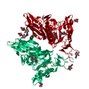
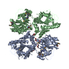
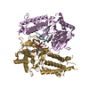
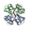
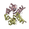
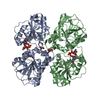
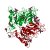
| Title | Crystal structure of rat coronavirus strain New-Jersey Hemagglutinin-Esterase in complex with 4N-acetyl sialic acid |
|---|
 Components Components | Hemagglutinin-esterase Hemagglutinin esterase Hemagglutinin esterase |
|---|
 Keywords Keywords | VIRAL PROTEIN / Hemagglutin / Esterase / Hepatitis virus / Coronavirus / sialic acid |
|---|
| Function / homology |  Function and homology information Function and homology information |
|---|
| Biological species |  Rat coronavirus Rat coronavirus |
|---|
| Method | X-RAY DIFFRACTION / SYNCHROTRON / MOLECULAR REPLACEMENT / Resolution: 1.85 Å |
|---|
 Authors Authors | Bakkers, M.J.G. / Feitsma, L.J. / de Groot, R.J. / Huizinga, E.G. |
|---|
| Funding support |  Netherlands, 1items Netherlands, 1items | Organization | Grant number | Country |
|---|
| Netherlands Organization for Scientific Research | ECHO 711.011.006 | Netherlands |
|
|---|
 Citation Citation | Journal: Proc.Natl.Acad.Sci.USA / Year: 2016
Title: Coronavirus receptor switch explained from the stereochemistry of protein-carbohydrate interactions and a single mutation.
Authors: Bakkers, M.J. / Zeng, Q. / Feitsma, L.J. / Hulswit, R.J. / Li, Z. / Westerbeke, A. / van Kuppeveld, F.J. / Boons, G.J. / Langereis, M.A. / Huizinga, E.G. / de Groot, R.J. |
|---|
| History | | Deposition | Apr 22, 2016 | Deposition site: RCSB / Processing site: PDBE |
|---|
| Revision 1.0 | May 11, 2016 | Provider: repository / Type: Initial release |
|---|
| Revision 1.1 | Jun 1, 2016 | Group: Database references |
|---|
| Revision 1.2 | Jun 8, 2016 | Group: Database references |
|---|
| Revision 1.3 | Sep 13, 2017 | Group: Author supporting evidence / Category: pdbx_audit_support / Item: _pdbx_audit_support.funding_organization |
|---|
| Revision 2.0 | Apr 3, 2019 | Group: Atomic model / Data collection / Source and taxonomy / Category: atom_site / entity_src_gen
Item: _atom_site.occupancy / _entity_src_gen.pdbx_host_org_cell_line |
|---|
| Revision 3.0 | Jul 29, 2020 | Group: Atomic model / Data collection ...Atomic model / Data collection / Derived calculations / Structure summary
Category: atom_site / atom_site_anisotrop ...atom_site / atom_site_anisotrop / chem_comp / entity / pdbx_branch_scheme / pdbx_chem_comp_identifier / pdbx_entity_branch / pdbx_entity_branch_descriptor / pdbx_entity_branch_link / pdbx_entity_branch_list / pdbx_entity_nonpoly / pdbx_nonpoly_scheme / pdbx_struct_assembly_gen / pdbx_struct_conn_angle / pdbx_struct_special_symmetry / struct_asym / struct_conn / struct_conn_type / struct_site / struct_site_gen
Item: _atom_site.B_iso_or_equiv / _atom_site.Cartn_x ..._atom_site.B_iso_or_equiv / _atom_site.Cartn_x / _atom_site.Cartn_y / _atom_site.Cartn_z / _atom_site.auth_asym_id / _atom_site.auth_atom_id / _atom_site.auth_seq_id / _atom_site.label_asym_id / _atom_site.label_atom_id / _atom_site.label_entity_id / _atom_site_anisotrop.U[1][1] / _atom_site_anisotrop.U[1][2] / _atom_site_anisotrop.U[1][3] / _atom_site_anisotrop.U[2][2] / _atom_site_anisotrop.U[2][3] / _atom_site_anisotrop.U[3][3] / _atom_site_anisotrop.pdbx_auth_asym_id / _atom_site_anisotrop.pdbx_auth_seq_id / _atom_site_anisotrop.pdbx_label_asym_id / _chem_comp.name / _chem_comp.type / _pdbx_entity_nonpoly.entity_id / _pdbx_entity_nonpoly.name / _pdbx_struct_assembly_gen.asym_id_list / _pdbx_struct_conn_angle.ptnr2_label_asym_id / _pdbx_struct_special_symmetry.label_asym_id / _struct_conn.conn_type_id / _struct_conn.id / _struct_conn.pdbx_dist_value / _struct_conn.pdbx_leaving_atom_flag / _struct_conn.pdbx_role / _struct_conn.ptnr1_auth_asym_id / _struct_conn.ptnr1_auth_comp_id / _struct_conn.ptnr1_auth_seq_id / _struct_conn.ptnr1_label_asym_id / _struct_conn.ptnr1_label_atom_id / _struct_conn.ptnr1_label_comp_id / _struct_conn.ptnr1_label_seq_id / _struct_conn.ptnr2_auth_asym_id / _struct_conn.ptnr2_auth_comp_id / _struct_conn.ptnr2_auth_seq_id / _struct_conn.ptnr2_label_asym_id / _struct_conn.ptnr2_label_atom_id / _struct_conn.ptnr2_label_comp_id / _struct_conn_type.id
Description: Carbohydrate remediation / Provider: repository / Type: Remediation |
|---|
| Revision 3.1 | May 1, 2024 | Group: Data collection / Database references ...Data collection / Database references / Refinement description / Structure summary
Category: chem_comp / chem_comp_atom ...chem_comp / chem_comp_atom / chem_comp_bond / database_2 / pdbx_initial_refinement_model
Item: _chem_comp.pdbx_synonyms / _database_2.pdbx_DOI / _database_2.pdbx_database_accession |
|---|
|
|---|
 Movie
Movie Controller

 Yorodumi
 Open data
 Basic information
 Structure visualization
 Downloads & links
 Other downloads
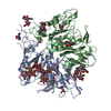

 PDBj
PDBj


 Assembly


 Type: D-saccharide, beta linking / Mass: 221.208 Da / Num. of mol.: 7
 Type: D-saccharide / Mass: 364.348 Da / Num. of mol.: 2 / Source method: obtained synthetically / Formula: C14H24N2O9


 Mass: 22.990 Da / Num. of mol.: 1 / Source method: obtained synthetically / Formula: Na
 Mass: 65.409 Da / Num. of mol.: 1 / Source method: obtained synthetically / Formula: Zn
 Sample preparation
 / Beamline: ID23-2 / Wavelength: 0.8729 Å
 Processing