 Movie
Movie Controller


+ Open data

- Basic information
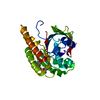
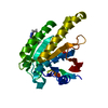
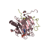
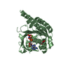
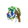
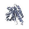
| Entry | Database: PDB / ID: 5jed | ||||||
|---|---|---|---|---|---|---|---|
| Title | Apo-structure of humanised RadA-mutant humRadA28 | ||||||
 Components Components | DNA repair and recombination protein RadA | ||||||
 Keywords Keywords |  HYDROLASE / DNA repair / fragment based drug design / humanisation HYDROLASE / DNA repair / fragment based drug design / humanisation | ||||||
| Function / homology |  Function and homology information Function and homology informationATP-dependent DNA damage sensor activity / DNA recombination / damaged DNA binding / DNA repair / ATP hydrolysis activity / ATP bindingSimilarity search - Function | ||||||
| Biological species |   Pyrococcus furiosus (archaea) Pyrococcus furiosus (archaea) | ||||||
| Method | X-RAY DIFFRACTION / SYNCHROTRON / MOLECULAR REPLACEMENT / Resolution: 1.332 Å | ||||||
 Authors Authors | Fischer, G. / Marsh, M. / Moschetti, T. / Sharpe, T. / Scott, D. / Morgan, M. / Ng, H. / Skidmore, J. / Venkitaraman, A. / Abell, C. ...Fischer, G. / Marsh, M. / Moschetti, T. / Sharpe, T. / Scott, D. / Morgan, M. / Ng, H. / Skidmore, J. / Venkitaraman, A. / Abell, C. / Blundell, T.L. / Hyvonen, M. | ||||||
 Citation Citation | Journal: J.Mol.Biol. / Year: 2016 Title: Engineering Archeal Surrogate Systems for the Development of Protein-Protein Interaction Inhibitors against Human RAD51. Authors: Moschetti, T. / Sharpe, T. / Fischer, G. / Marsh, M.E. / Ng, H.K. / Morgan, M. / Scott, D.E. / Blundell, T.L. / R Venkitaraman, A. / Skidmore, J. / Abell, C. / Hyvonen, M. | ||||||
| History |
|
- Structure visualization
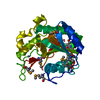
| Structure viewer | Molecule: MolmilJmol/JSmol |
|---|
- Downloads & links
-Download
| PDBx/mmCIF format | 5jed.cif.gz | 120.1 KB | Display | PDBx/mmCIF format |
|---|---|---|---|---|
| PDB format | pdb5jed.ent.gz | 91.8 KB | Display | PDB format |
| PDBx/mmJSON format | 5jed.json.gz | Tree view | PDBx/mmJSON format | |
| Others |  Other downloads Other downloads |
-Validation report
| Arichive directory | https://data.pdbj.org/pub/pdb/validation_reports/je/5jedftp://data.pdbj.org/pub/pdb/validation_reports/je/5jed | HTTPS FTP |
|---|
-Related structure data
| Related structure data | 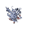 5fosC  5j4hC 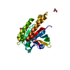 5j4kC 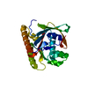 5j4lSC 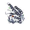 5jecC 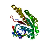 5jeeC  5kddC  5l8vC  5lb2C  5lb4C  5lbiC S: Starting model for refinement C: citing same article ( |
|---|---|
| Similar structure data |


-Links
 PDBj
PDBj


- Assembly
| Deposited unit | 
| ||||||||
|---|---|---|---|---|---|---|---|---|---|
| 1 |
| ||||||||
| Unit cell |
|
-Components
-Protein , 1 types, 1 molecules A
| #1: Protein | Mass: 25628.178 Da / Num. of mol.: 1 Mutation: S167K, V168A, I169M, W170Y, I182L, K198D, H199N, I200V, Y201A, V202Y, L213Q, V215L, Q216Y, E219S, K221M, I222M, K223V, L225S, V232Y, K263R, H264F, A266R, D267M, L274E, Y275F Source method: isolated from a genetically manipulated source Source: (gene. exp.) Pyrococcus furiosus (archaea) / Strain: ATCC 43587 / DSM 3638 / JCM 8422 / Vc1 / Gene: radA, PF1926 / Plasmid: pBAT4 / Production host:  Escherichia coli BL21(DE3) (bacteria) / References: UniProt: O74036 Escherichia coli BL21(DE3) (bacteria) / References: UniProt: O74036 |
|---|
-Non-polymers , 5 types, 205 molecules 


| #2: Chemical | ChemComp-SO4 / Sulfate Mass: 96.063 Da / Num. of mol.: 1 / Source method: obtained synthetically / Formula: SO4 Mass: 96.063 Da / Num. of mol.: 1 / Source method: obtained synthetically / Formula: SO4 |
|---|---|
| #3: Chemical | ChemComp-MPD / (2-Methyl-2,4-pentanediol Mass: 118.174 Da / Num. of mol.: 1 / Source method: obtained synthetically / Formula: C6H14O2 / Comment: precipitant*YM Mass: 118.174 Da / Num. of mol.: 1 / Source method: obtained synthetically / Formula: C6H14O2 / Comment: precipitant*YM |
| #4: Chemical | ChemComp-MRD / (2-Methyl-2,4-pentanediol Mass: 118.174 Da / Num. of mol.: 1 / Source method: obtained synthetically / Formula: C6H14O2 / Comment: precipitant*YM Mass: 118.174 Da / Num. of mol.: 1 / Source method: obtained synthetically / Formula: C6H14O2 / Comment: precipitant*YM |
| #5: Chemical | ChemComp-CL / Chloride Mass: 35.453 Da / Num. of mol.: 1 / Source method: obtained synthetically / Formula: Cl Mass: 35.453 Da / Num. of mol.: 1 / Source method: obtained synthetically / Formula: Cl |
| #6: Water | ChemComp-HOH / WaterMass: 18.015 Da / Num. of mol.: 201 / Source method: isolated from a natural source / Formula: H2O |
-Experimental details
-Experiment
| Experiment | Method: X-RAY DIFFRACTION / Number of used crystals: 1 |
|---|
- Sample preparation
| Crystal | Density Matthews: 2 Å3/Da / Density % sol: 38.62 % / Description: rods |
|---|---|
| Crystal grow | Temperature: 292 K / Method: vapor diffusion, hanging drop / pH: 6.5 / Details: 0.1M NaCacodylate pH=6.5, 5% PEG8000, 40% MPD |
-Data collection
| Diffraction | Mean temperature: 100 K |
|---|---|
| Diffraction source | Source: SYNCHROTRON / Site: Diamond  / Beamline: I24 / Wavelength: 0.96862 Å / Beamline: I24 / Wavelength: 0.96862 Å |
| Detector | Type: DECTRIS PILATUS 6M / Detector: PIXEL / Date: Feb 13, 2012 |
| Radiation | Protocol: SINGLE WAVELENGTH / Monochromatic (M) / Laue (L): M / Scattering type: x-ray |
| Radiation wavelength | Wavelength: 0.96862 Å / Relative weight: 1 |
| Reflection | Resolution: 1.332→37.72 Å / Num. obs: 45801 / % possible obs: 99.3 % / Redundancy: 4.7 % / CC1/2: 0.992 / Rmerge(I) obs: 0.093 / Rsym value: 0.103 / Net I/σ(I): 7.6 |
| Reflection shell | Resolution: 1.332→1.337 Å / Redundancy: 3.2 % / Rmerge(I) obs: 1.086 / Mean I/σ(I) obs: 2.1 / % possible all: 99.1 |
- Processing
| Software |
| ||||||||||||||||||||||||||||||||||||||||||||||||||||||||||||||||||||||||||||||||||||||||||||||||||||||||||||||||||||||||||||||||||||||||||||||||||||||||||||||||||||||||||||||||||||||
|---|---|---|---|---|---|---|---|---|---|---|---|---|---|---|---|---|---|---|---|---|---|---|---|---|---|---|---|---|---|---|---|---|---|---|---|---|---|---|---|---|---|---|---|---|---|---|---|---|---|---|---|---|---|---|---|---|---|---|---|---|---|---|---|---|---|---|---|---|---|---|---|---|---|---|---|---|---|---|---|---|---|---|---|---|---|---|---|---|---|---|---|---|---|---|---|---|---|---|---|---|---|---|---|---|---|---|---|---|---|---|---|---|---|---|---|---|---|---|---|---|---|---|---|---|---|---|---|---|---|---|---|---|---|---|---|---|---|---|---|---|---|---|---|---|---|---|---|---|---|---|---|---|---|---|---|---|---|---|---|---|---|---|---|---|---|---|---|---|---|---|---|---|---|---|---|---|---|---|---|---|---|---|---|
| Refinement | Method to determine structure: MOLECULAR REPLACEMENT Starting model: 5j4l Resolution: 1.332→37.72 Å / Cor.coef. Fo:Fc: 0.98 / Cor.coef. Fo:Fc free: 0.971 / SU B: 2.353 / SU ML: 0.041 / Cross valid method: THROUGHOUT / ESU R: 0.055 / ESU R Free: 0.052 / Details: HYDROGENS HAVE BEEN ADDED IN THE RIDING POSITIONS
| ||||||||||||||||||||||||||||||||||||||||||||||||||||||||||||||||||||||||||||||||||||||||||||||||||||||||||||||||||||||||||||||||||||||||||||||||||||||||||||||||||||||||||||||||||||||
| Solvent computation | Ion probe radii: 0.8 Å / Shrinkage radii: 0.8 Å / VDW probe radii: 1.2 Å | ||||||||||||||||||||||||||||||||||||||||||||||||||||||||||||||||||||||||||||||||||||||||||||||||||||||||||||||||||||||||||||||||||||||||||||||||||||||||||||||||||||||||||||||||||||||
| Displacement parameters | Biso mean: 21.216 Å2
| ||||||||||||||||||||||||||||||||||||||||||||||||||||||||||||||||||||||||||||||||||||||||||||||||||||||||||||||||||||||||||||||||||||||||||||||||||||||||||||||||||||||||||||||||||||||
| Refinement step | Cycle: 1 / Resolution: 1.332→37.72 Å
| ||||||||||||||||||||||||||||||||||||||||||||||||||||||||||||||||||||||||||||||||||||||||||||||||||||||||||||||||||||||||||||||||||||||||||||||||||||||||||||||||||||||||||||||||||||||
| Refine LS restraints |
|