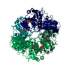
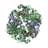
Entry Database : PDB / ID : 5j9gTitle Structure of Lactobacillus acidophilus glyceraldehyde-3-phosphate dehydrogenase at 2.21 angstrom resolution Glyceraldehyde-3-p dehydrogenase Keywords / / Function / homology Function Domain/homology Component
/ / / / / / / / / / / / / / / / / / / / / / / Biological species Lactobacillus acidophilus (bacteria)Method / / Resolution : 2.21 Å Authors Patel, D. / Pappachan, A. / Singh, D.D. Funding support Organization Grant number Country Department of Biotechnology
Journal : To Be Published Title : Structure of Lactobacillus acidophilus glyceraldehyde-3-phosphate dehydrogenase at 2.21 angstrom resolution.Authors : Patel, D. / Pappachan, A. / Singh, D.D. History Deposition Apr 10, 2016 Deposition site / Processing site Revision 1.0 Apr 19, 2017 Provider / Type Revision 1.1 Nov 8, 2023 Group / Database references / Refinement descriptionCategory chem_comp_atom / chem_comp_bond ... chem_comp_atom / chem_comp_bond / database_2 / pdbx_initial_refinement_model / struct_ncs_dom_lim Item _database_2.pdbx_DOI / _database_2.pdbx_database_accession ... _database_2.pdbx_DOI / _database_2.pdbx_database_accession / _struct_ncs_dom_lim.beg_auth_comp_id / _struct_ncs_dom_lim.beg_label_asym_id / _struct_ncs_dom_lim.beg_label_comp_id / _struct_ncs_dom_lim.beg_label_seq_id / _struct_ncs_dom_lim.end_auth_comp_id / _struct_ncs_dom_lim.end_label_asym_id / _struct_ncs_dom_lim.end_label_comp_id / _struct_ncs_dom_lim.end_label_seq_id
Show all Show less
 Movie
Movie Controller

 Yorodumi
 Open data
 Basic information
 Components
 Keywords
 OXIDOREDUCTASE /
  Function and homology information
 Authors
 India, 1items
  Citation
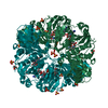
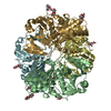
 Structure visualization
 Downloads & links
 Other downloads


 PDBj
PDBj
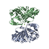
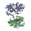
 Assembly
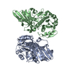

 Mass: 18.015 Da / Num. of mol.: 89 / Source method: isolated from a natural source / Formula: H2O
 Sample preparation
 Processing