 Movie
Movie Controller


+ Open data

- Basic information
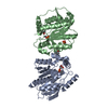
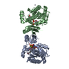
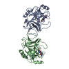
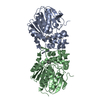
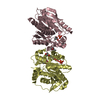
| Entry | Database: PDB / ID: 5j8j | ||||||
|---|---|---|---|---|---|---|---|
| Title | A histone deacetylase from Saccharomyces cerevisiae | ||||||
 Components Components | Histone deacetylase HDA1 | ||||||
 Keywords Keywords |  HYDROLASE / Rossmann fold / deacetylase HYDROLASE / Rossmann fold / deacetylase | ||||||
| Function / homology |  Function and homology information Function and homology informationHDA1 complex / HDACs deacetylate histones / negative regulation of transcription by transcription factor localization / HSF1 activation / SUMOylation of chromatin organization proteins / histone deacetylase / histone deacetylase activity / histone deacetylase complex / chromatin binding / negative regulation of transcription by RNA polymerase II ...HDA1 complex / HDACs deacetylate histones / negative regulation of transcription by transcription factor localization / HSF1 activation / SUMOylation of chromatin organization proteins / histone deacetylase / histone deacetylase activity / histone deacetylase complex / chromatin binding / negative regulation of transcription by RNA polymerase II / positive regulation of transcription by RNA polymerase II / identical protein binding / cytoplasmSimilarity search - Function | ||||||
| Biological species |  Saccharomyces cerevisiae (brewer's yeast) Saccharomyces cerevisiae (brewer's yeast) | ||||||
| Method | X-RAY DIFFRACTION / SYNCHROTRON / SAD / Resolution: 2.716 Å | ||||||
 Authors Authors | Zhu, Y. / Shen, H. / Li, X. / Teng, M. | ||||||
 Citation Citation | Journal: Sci Rep / Year: 2016 Title: Structural and histone binding ability characterization of the ARB2 domain of a histone deacetylase Hda1 from Saccharomyces cerevisiae. Authors: Shen, H. / Zhu, Y. / Wang, C. / Yan, H. / Teng, M. / Li, X. | ||||||
| History |
|
- Structure visualization
| Structure viewer | Molecule: MolmilJmol/JSmol |
|---|
- Downloads & links
-Download
| PDBx/mmCIF format | 5j8j.cif.gz | 106.6 KB | Display | PDBx/mmCIF format |
|---|---|---|---|---|
| PDB format | pdb5j8j.ent.gz | 86.6 KB | Display | PDB format |
| PDBx/mmJSON format | 5j8j.json.gz | Tree view | PDBx/mmJSON format | |
| Others |  Other downloads Other downloads |
-Validation report
| Arichive directory | https://data.pdbj.org/pub/pdb/validation_reports/j8/5j8jftp://data.pdbj.org/pub/pdb/validation_reports/j8/5j8j | HTTPS FTP |
|---|
-Related structure data
| Similar structure data |
|---|


-Links
 PDBj
PDBj
- Assembly
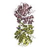
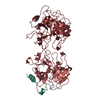
| Deposited unit | 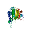
| ||||||||
|---|---|---|---|---|---|---|---|---|---|
| 1 | 
| ||||||||
| Unit cell |
|
-Components
| #1: Protein | Mass: 28885.723 Da / Num. of mol.: 1 / Fragment: UNP RESIDUES 457-698 Source method: isolated from a genetically manipulated source Source: (gene. exp.) Saccharomyces cerevisiae (strain ATCC 204508 / S288c) (yeast)Strain: ATCC 204508 / S288c / Gene: HDA1, YNL021W, N2819 / Production host:  Escherichia coli (E. coli) / References: UniProt: P53973, histone deacetylase Escherichia coli (E. coli) / References: UniProt: P53973, histone deacetylase |
|---|
-Experimental details
-Experiment
| Experiment | Method: X-RAY DIFFRACTION / Number of used crystals: 1 |
|---|
- Sample preparation
| Crystal | Density Matthews: 4.79 Å3/Da / Density % sol: 74.31 % |
|---|---|
| Crystal grow | Temperature: 285 K / Method: vapor diffusion, hanging drop / pH: 4.6 Details: 0.1 M Sodium acetate trihydrate pH 4.6, 2.0 M Sodium formate |
-Data collection
| Diffraction | Mean temperature: 100 K |
|---|---|
| Diffraction source | Source: SYNCHROTRON / Site: APS  / Beamline: 17-BM / Wavelength: 0.9792 Å / Beamline: 17-BM / Wavelength: 0.9792 Å |
| Detector | Type: MAR CCD 130 mm / Detector: CCD / Date: Mar 22, 2016 |
| Radiation | Protocol: SINGLE WAVELENGTH / Monochromatic (M) / Laue (L): M / Scattering type: x-ray |
| Radiation wavelength | Wavelength: 0.9792 Å / Relative weight: 1 |
| Reflection | Resolution: 2.7→50 Å / Num. obs: 14972 / % possible obs: 98.1 % / Redundancy: 10 % / Rmerge(I) obs: 0.086 / Net I/σ(I): 14.7 |
| Reflection shell | Redundancy: 10.3 % / Mean I/σ(I) obs: 3.1 / % possible all: 96.9 |
- Processing
| Software |
| |||||||||||||||||||||||||||||||||||||||||||||||||
|---|---|---|---|---|---|---|---|---|---|---|---|---|---|---|---|---|---|---|---|---|---|---|---|---|---|---|---|---|---|---|---|---|---|---|---|---|---|---|---|---|---|---|---|---|---|---|---|---|---|---|
| Refinement | Method to determine structure: SAD / Resolution: 2.716→40.284 Å / SU ML: 0.37 / Cross valid method: FREE R-VALUE / σ(F): 1.36 / Phase error: 29.82
| |||||||||||||||||||||||||||||||||||||||||||||||||
| Solvent computation | Shrinkage radii: 0.9 Å / VDW probe radii: 1.11 Å | |||||||||||||||||||||||||||||||||||||||||||||||||
| Refinement step | Cycle: LAST / Resolution: 2.716→40.284 Å
| |||||||||||||||||||||||||||||||||||||||||||||||||
| Refine LS restraints |
| |||||||||||||||||||||||||||||||||||||||||||||||||
| LS refinement shell |
| |||||||||||||||||||||||||||||||||||||||||||||||||
| Refinement TLS params. | Method: refined / Origin x: -46.2438 Å / Origin y: 18.2399 Å / Origin z: -25.4993 Å
| |||||||||||||||||||||||||||||||||||||||||||||||||
| Refinement TLS group | Selection details: all |
