 Movie
Movie Controller

[English] 日本語
 Yorodumi
- PDB-5j81: Crystal structure of Glycoprotein C from Puumala virus in the pos... -
+ Open data

- Basic information
| Entry | Database: PDB / ID: 5j81 | |||||||||
|---|---|---|---|---|---|---|---|---|---|---|
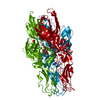
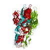
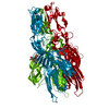
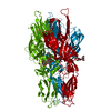
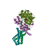
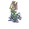
| Title | Crystal structure of Glycoprotein C from Puumala virus in the post-fusion conformation (pH 6.0) | |||||||||
 Components Components | Envelopment polyprotein | |||||||||
 Keywords Keywords |  VIRAL PROTEIN / membrane fusion / virus / glycoprotein / class II VIRAL PROTEIN / membrane fusion / virus / glycoprotein / class II | |||||||||
| Function / homology |  Function and homology information Function and homology information: / symbiont-mediated suppression of host TRAF-mediated signal transduction / host cell Golgi membrane / host cell mitochondrion / host cell surface / host cell endoplasmic reticulum membrane / symbiont entry into host cell / fusion of virus membrane with host endosome membrane / virion attachment to host cell / virion membrane ...: / symbiont-mediated suppression of host TRAF-mediated signal transduction / host cell Golgi membrane / host cell mitochondrion / host cell surface / host cell endoplasmic reticulum membrane / symbiont entry into host cell / fusion of virus membrane with host endosome membrane / virion attachment to host cell / virion membrane / signal transduction / membrane / metal ion bindingSimilarity search - Function | |||||||||
| Biological species |  Puumala virus Puumala virus | |||||||||
| Method | X-RAY DIFFRACTION / SYNCHROTRON / SIRAS / Resolution: 1.8 Å | |||||||||
 Authors Authors | Willensky, S. / Dessau, M. | |||||||||
 Citation Citation | Journal: Plos Pathog. / Year: 2016 Title: Crystal Structure of Glycoprotein C from a Hantavirus in the Post-fusion Conformation. Authors: Willensky, S. / Bar-Rogovsky, H. / Bignon, E.A. / Tischler, N.D. / Modis, Y. / Dessau, M. | |||||||||
| History |
|
- Structure visualization
| Structure viewer | Molecule: MolmilJmol/JSmol |
|---|
- Downloads & links
-Download
| PDBx/mmCIF format | 5j81.cif.gz | 178.4 KB | Display | PDBx/mmCIF format |
|---|---|---|---|---|
| PDB format | pdb5j81.ent.gz | 141.9 KB | Display | PDB format |
| PDBx/mmJSON format | 5j81.json.gz | Tree view | PDBx/mmJSON format | |
| Others |  Other downloads Other downloads |
-Validation report
| Arichive directory | https://data.pdbj.org/pub/pdb/validation_reports/j8/5j81ftp://data.pdbj.org/pub/pdb/validation_reports/j8/5j81 | HTTPS FTP |
|---|
-Related structure data


-Links
 PDBj
PDBj- Assembly
| Deposited unit | 
| ||||||||||||
|---|---|---|---|---|---|---|---|---|---|---|---|---|---|
| 1 | 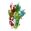
| ||||||||||||
| Unit cell |
| ||||||||||||
| Components on special symmetry positions |
|
-Components
-Protein / Sugars , 2 types, 2 molecules A
| #1: Protein | Mass: 48986.305 Da / Num. of mol.: 1 Source method: isolated from a genetically manipulated source Source: (gene. exp.) Puumala virus (strain P360) / Strain: P360 / Gene: GP / Production host:  Trichoplusia ni (cabbage looper) / References: UniProt: P41266 Trichoplusia ni (cabbage looper) / References: UniProt: P41266 |
|---|---|
| #2: Polysaccharide | beta-D-mannopyranose-(1-4)-2-acetamido-2-deoxy-beta-D-glucopyranose-(1-4)-2-acetamido-2-deoxy-beta- ...beta-D-mannopyranose-(1-4)-2-acetamido-2-deoxy-beta-D-glucopyranose-(1-4)-2-acetamido-2-deoxy-beta-D-glucopyranose / Mass: 586.542 Da / Num. of mol.: 1 Source method: isolated from a genetically manipulated source |
-Non-polymers , 4 types, 370 molecules 


| #3: Chemical | ChemComp-P33 / Polyethylene glycol Mass: 326.383 Da / Num. of mol.: 1 / Source method: obtained synthetically / Formula: C14H30O8 / Comment: precipitant*YM Mass: 326.383 Da / Num. of mol.: 1 / Source method: obtained synthetically / Formula: C14H30O8 / Comment: precipitant*YM |
|---|---|
| #4: Chemical | ChemComp-SO4 / Sulfate Mass: 96.063 Da / Num. of mol.: 1 / Source method: obtained synthetically / Formula: SO4 Mass: 96.063 Da / Num. of mol.: 1 / Source method: obtained synthetically / Formula: SO4 |
| #5: Chemical | ChemComp-ACT / Acetate Mass: 59.044 Da / Num. of mol.: 1 / Source method: obtained synthetically / Formula: C2H3O2 Mass: 59.044 Da / Num. of mol.: 1 / Source method: obtained synthetically / Formula: C2H3O2 |
| #6: Water | ChemComp-HOH / WaterMass: 18.015 Da / Num. of mol.: 367 / Source method: isolated from a natural source / Formula: H2O |
-Experimental details
-Experiment
| Experiment | Method: X-RAY DIFFRACTION / Number of used crystals: 1 |
|---|
- Sample preparation
| Crystal | Density Matthews: 2.46 Å3/Da / Density % sol: 50.09 % / Description: Sharp edges rhombohedrons |
|---|---|
| Crystal grow | Temperature: 289 K / Method: vapor diffusion, hanging drop / pH: 6 Details: 12% PEG 2000 MME, 0.1 M MES pH 6.0, 0.2 M (NH4)2SO4 |
-Data collection
| Diffraction | Mean temperature: 100 K | ||||||||||||||||||||||||||||||||||||||||||||||||||||||||||||||||||||||||||||||||||||||||||||||||||||||
|---|---|---|---|---|---|---|---|---|---|---|---|---|---|---|---|---|---|---|---|---|---|---|---|---|---|---|---|---|---|---|---|---|---|---|---|---|---|---|---|---|---|---|---|---|---|---|---|---|---|---|---|---|---|---|---|---|---|---|---|---|---|---|---|---|---|---|---|---|---|---|---|---|---|---|---|---|---|---|---|---|---|---|---|---|---|---|---|---|---|---|---|---|---|---|---|---|---|---|---|---|---|---|---|
| Diffraction source | Source: SYNCHROTRON / Site: Diamond  / Beamline: I04 / Wavelength: 0.97949 Å / Beamline: I04 / Wavelength: 0.97949 Å | ||||||||||||||||||||||||||||||||||||||||||||||||||||||||||||||||||||||||||||||||||||||||||||||||||||||
| Detector | Type: DECTRIS PILATUS3 6M / Detector: PIXEL / Date: Sep 28, 2015 | ||||||||||||||||||||||||||||||||||||||||||||||||||||||||||||||||||||||||||||||||||||||||||||||||||||||
| Radiation | Protocol: SINGLE WAVELENGTH / Monochromatic (M) / Laue (L): M / Scattering type: x-ray | ||||||||||||||||||||||||||||||||||||||||||||||||||||||||||||||||||||||||||||||||||||||||||||||||||||||
| Radiation wavelength | Wavelength: 0.97949 Å / Relative weight: 1 | ||||||||||||||||||||||||||||||||||||||||||||||||||||||||||||||||||||||||||||||||||||||||||||||||||||||
| Reflection | Resolution: 1.8→50 Å / Num. obs: 41365 / % possible obs: 99.9 % / Observed criterion σ(I): -3 / Redundancy: 9.93 % / CC1/2: 0.999 / Rmerge(I) obs: 0.094 / Net I/σ(I): 14.65 | ||||||||||||||||||||||||||||||||||||||||||||||||||||||||||||||||||||||||||||||||||||||||||||||||||||||
| Reflection shell |
|
- Processing
| Software |
| |||||||||||||||||||||||||||||||||||||||||||||||||||||||||||||||||||||||||||||||||||||||||||||||||||||||||||||||||||||||||||||
|---|---|---|---|---|---|---|---|---|---|---|---|---|---|---|---|---|---|---|---|---|---|---|---|---|---|---|---|---|---|---|---|---|---|---|---|---|---|---|---|---|---|---|---|---|---|---|---|---|---|---|---|---|---|---|---|---|---|---|---|---|---|---|---|---|---|---|---|---|---|---|---|---|---|---|---|---|---|---|---|---|---|---|---|---|---|---|---|---|---|---|---|---|---|---|---|---|---|---|---|---|---|---|---|---|---|---|---|---|---|---|---|---|---|---|---|---|---|---|---|---|---|---|---|---|---|---|
| Refinement | Method to determine structure: SIRAS / Resolution: 1.8→48.215 Å / SU ML: 0.26 / Cross valid method: FREE R-VALUE / σ(F): 1.34 / Phase error: 21.26
| |||||||||||||||||||||||||||||||||||||||||||||||||||||||||||||||||||||||||||||||||||||||||||||||||||||||||||||||||||||||||||||
| Solvent computation | Shrinkage radii: 0.9 Å / VDW probe radii: 1.11 Å | |||||||||||||||||||||||||||||||||||||||||||||||||||||||||||||||||||||||||||||||||||||||||||||||||||||||||||||||||||||||||||||
| Displacement parameters | Biso max: 122.99 Å2 / Biso mean: 25.3894 Å2 / Biso min: 4.24 Å2 | |||||||||||||||||||||||||||||||||||||||||||||||||||||||||||||||||||||||||||||||||||||||||||||||||||||||||||||||||||||||||||||
| Refinement step | Cycle: final / Resolution: 1.8→48.215 Å
| |||||||||||||||||||||||||||||||||||||||||||||||||||||||||||||||||||||||||||||||||||||||||||||||||||||||||||||||||||||||||||||
| Refine LS restraints |
| |||||||||||||||||||||||||||||||||||||||||||||||||||||||||||||||||||||||||||||||||||||||||||||||||||||||||||||||||||||||||||||
| LS refinement shell | Refine-ID: X-RAY DIFFRACTION / Total num. of bins used: 15 / % reflection obs: 100 %
| |||||||||||||||||||||||||||||||||||||||||||||||||||||||||||||||||||||||||||||||||||||||||||||||||||||||||||||||||||||||||||||
| Refinement TLS params. | Method: refined / Refine-ID: X-RAY DIFFRACTION
| |||||||||||||||||||||||||||||||||||||||||||||||||||||||||||||||||||||||||||||||||||||||||||||||||||||||||||||||||||||||||||||
| Refinement TLS group |
|
