 Movie
Movie Controller

[English] 日本語
 Yorodumi
- PDB-5j6o: Crystal structure of a trans-AT PKS dehydratase domain of C0ZGQ7 ... -
+ Open data

- Basic information
| Entry | Database: PDB / ID: 5j6o | ||||||
|---|---|---|---|---|---|---|---|
| Title | Crystal structure of a trans-AT PKS dehydratase domain of C0ZGQ7 from Brevibacillus brevis | ||||||
 Components Components | Putative polyketide synthase | ||||||
 Keywords Keywords | LYASE / polyketide / trans-AT / PKS | ||||||
| Function / homology |  Function and homology informationphosphopantetheine binding / 3-oxoacyl-[acyl-carrier-protein] synthase activity / fatty acid biosynthetic process Function and homology informationphosphopantetheine binding / 3-oxoacyl-[acyl-carrier-protein] synthase activity / fatty acid biosynthetic processSimilarity search - Function | ||||||
| Biological species |  Brevibacillus brevis (bacteria) Brevibacillus brevis (bacteria) | ||||||
| Method | X-RAY DIFFRACTION / SYNCHROTRON / MOLECULAR REPLACEMENT / Resolution: 3.195 Å | ||||||
 Authors Authors | Jakob, R.P. / Herbst, D.A. / Maier, T. | ||||||
 Citation Citation | Journal: To Be Published Title: Crystal Structures of Dehydratase Domains from trans-AT Polyketide Biosynthetic Pathway Authors: Jakob, R.P. / Herbst, D.A. / Muller, R. / Maier, T. | ||||||
| History |
|
- Structure visualization
| Structure viewer | Molecule: MolmilJmol/JSmol |
|---|
- Downloads & links
-Download
| PDBx/mmCIF format | 5j6o.cif.gz | 113.9 KB | Display | PDBx/mmCIF format |
|---|---|---|---|---|
| PDB format | pdb5j6o.ent.gz | 88.9 KB | Display | PDB format |
| PDBx/mmJSON format | 5j6o.json.gz | Tree view | PDBx/mmJSON format | |
| Others |  Other downloads Other downloads |
-Validation report
| Arichive directory | https://data.pdbj.org/pub/pdb/validation_reports/j6/5j6oftp://data.pdbj.org/pub/pdb/validation_reports/j6/5j6o | HTTPS FTP |
|---|
-Related structure data
| Related structure data |  5hstC  5hu7C  5il6C  3kg9S S: Starting model for refinement C: citing same article ( |
|---|---|
| Similar structure data |


-Links
 PDBj
PDBj

- Assembly
| Deposited unit | 
| ||||||||
|---|---|---|---|---|---|---|---|---|---|
| 1 | 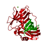
| ||||||||
| Unit cell |
| ||||||||
| Components on special symmetry positions |
|
-Components
| #1: Protein | Mass: 35397.941 Da / Num. of mol.: 1 / Fragment: UNP residues 1922-2240 Source method: isolated from a genetically manipulated source Source: (gene. exp.) Brevibacillus brevis (strain 47 / JCM 6285 / NBRC 100599) (bacteria)Strain: 47 / JCM 6285 / NBRC 100599 / Gene: BBR47_39890 / Plasmid: pNIC28a-Bsa4 / Production host: Escherichia coli BL21(DE3) (bacteria) / References: UniProt: C0ZGQ7 |
|---|---|
| #2: Chemical | ChemComp-MG /   Mass: 24.305 Da / Num. of mol.: 1 / Source method: obtained synthetically / Formula: Mg Mass: 24.305 Da / Num. of mol.: 1 / Source method: obtained synthetically / Formula: Mg |
| #3: Water | ChemComp-HOH / Water Mass: 18.015 Da / Num. of mol.: 5 / Source method: isolated from a natural source / Formula: H2O Mass: 18.015 Da / Num. of mol.: 5 / Source method: isolated from a natural source / Formula: H2O |
-Experimental details
-Experiment
| Experiment | Method: X-RAY DIFFRACTION / Number of used crystals: 1 |
|---|
- Sample preparation
| Crystal | Density Matthews: 2.07 Å3/Da / Density % sol: 40.52 % |
|---|---|
| Crystal grow | Temperature: 293 K / Method: vapor diffusion, sitting drop / pH: 7.5 Details: 10% PEG20000, 20% PEG550MME, 0.1 M Mops/Hepes pH 7.5, 0.1 M MgCl2 |
-Data collection
| Diffraction | Mean temperature: 100 K |
|---|---|
| Diffraction source | Source: SYNCHROTRON / Site: SLS  / Beamline: X06SA / Wavelength: 1 Å / Beamline: X06SA / Wavelength: 1 Å |
| Detector | Type: DECTRIS PILATUS3 6M / Detector: PIXEL / Date: Feb 18, 2015 |
| Radiation | Protocol: SINGLE WAVELENGTH / Monochromatic (M) / Laue (L): M / Scattering type: x-ray |
| Radiation wavelength | Wavelength: 1 Å / Relative weight: 1 |
| Reflection | Resolution: 3.195→47.8 Å / Num. obs: 5296 / % possible obs: 95.5 % / Redundancy: 6.5 % / CC1/2: 0.99 / Rmerge(I) obs: 0.062 / Net I/σ(I): 21.6 |
| Reflection shell | Resolution: 3.195→3.3 Å / Redundancy: 6.3 % / Rmerge(I) obs: 0.885 / Mean I/σ(I) obs: 2.2 / % possible all: 93.1 |
- Processing
| Software |
| |||||||||||||||||||||||||||||||||||
|---|---|---|---|---|---|---|---|---|---|---|---|---|---|---|---|---|---|---|---|---|---|---|---|---|---|---|---|---|---|---|---|---|---|---|---|---|
| Refinement | Method to determine structure: MOLECULAR REPLACEMENT Starting model: 3KG9 Resolution: 3.195→47.8 Å / SU ML: 0.57 / Cross valid method: FREE R-VALUE / σ(F): 1.36 / Phase error: 37.82
| |||||||||||||||||||||||||||||||||||
| Solvent computation | Shrinkage radii: 0.9 Å / VDW probe radii: 1.11 Å | |||||||||||||||||||||||||||||||||||
| Refinement step | Cycle: LAST / Resolution: 3.195→47.8 Å
| |||||||||||||||||||||||||||||||||||
| Refine LS restraints |
| |||||||||||||||||||||||||||||||||||
| LS refinement shell |
|
