 Movie
Movie Controller

[English] 日本語
 Yorodumi
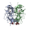
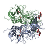
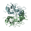
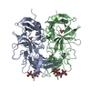
- PDB-5j3o: Protruding domain of GII.4 human norovirus NSW0514 in complex wit... -
+ Open data

- Basic information
| Entry | Database: PDB / ID: 5j3o | ||||||
|---|---|---|---|---|---|---|---|
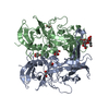
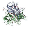
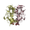
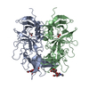
| Title | Protruding domain of GII.4 human norovirus NSW0514 in complex with 3-fucosyllactose (3FL) | ||||||
 Components Components | VP1 | ||||||
 Keywords Keywords |  VIRAL PROTEIN / Viral capsid protein / Protruding domain / Norovirus / Human milk oligosaccharides VIRAL PROTEIN / Viral capsid protein / Protruding domain / Norovirus / Human milk oligosaccharides | ||||||
| Function / homology |  Function and homology information Function and homology informationPositive stranded ssRNA viruses / Nucleoplasmin-like/VP (viral coat and capsid proteins) / Positive stranded ssRNA viruses / Calicivirus coat protein C-terminal / Calicivirus coat protein C-terminal / Calicivirus coat protein / Calicivirus coat protein / Elongation Factor Tu (Ef-tu); domain 3 / Picornavirus/Calicivirus coat protein / Viral coat protein subunit ...Positive stranded ssRNA viruses / Nucleoplasmin-like/VP (viral coat and capsid proteins) / Positive stranded ssRNA viruses / Calicivirus coat protein C-terminal / Calicivirus coat protein C-terminal / Calicivirus coat protein / Calicivirus coat protein / Elongation Factor Tu (Ef-tu); domain 3 / Picornavirus/Calicivirus coat protein / Viral coat protein subunit / Beta Barrel / Mainly BetaSimilarity search - Domain/homology | ||||||
| Biological species |  Norovirus Hu/GII.4/Sydney/NSW0514/2012/AU Norovirus Hu/GII.4/Sydney/NSW0514/2012/AU | ||||||
| Method | X-RAY DIFFRACTION / SYNCHROTRON / MOLECULAR REPLACEMENT / Resolution: 1.47 Å | ||||||
 Authors Authors | Singh, B.K. / Hansman, G.S. | ||||||
 Citation Citation | Journal: To be published Title: Structural basis for GII.4 norovirus inhibition by human milk oligosaccharides. Authors: Singh, B.K. / Hansman, G.S. | ||||||
| History |
|
- Structure visualization
| Structure viewer | Molecule: MolmilJmol/JSmol |
|---|
- Downloads & links
-Download
| PDBx/mmCIF format | 5j3o.cif.gz | 360.1 KB | Display | PDBx/mmCIF format |
|---|---|---|---|---|
| PDB format | pdb5j3o.ent.gz | 298.1 KB | Display | PDB format |
| PDBx/mmJSON format | 5j3o.json.gz | Tree view | PDBx/mmJSON format | |
| Others |  Other downloads Other downloads |
-Validation report
| Arichive directory | https://data.pdbj.org/pub/pdb/validation_reports/j3/5j3oftp://data.pdbj.org/pub/pdb/validation_reports/j3/5j3o | HTTPS FTP |
|---|
-Related structure data
| Related structure data | 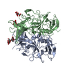 5iyrC 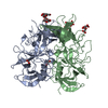 5iywC  5j35C  4oosS C: citing same article ( S: Starting model for refinement |
|---|---|
| Similar structure data |


-Links
 PDBj
PDBj

- Assembly
| Deposited unit | 
| ||||||||
|---|---|---|---|---|---|---|---|---|---|
| 1 |
| ||||||||
| Unit cell |
|
-Components
| #1: Protein | Mass: 34215.129 Da / Num. of mol.: 2 Source method: isolated from a genetically manipulated source Source: (gene. exp.) Norovirus Hu/GII.4/Sydney/NSW0514/2012/AUPlasmid: MBP-HTSHP / Production host:  Escherichia coli BL21(DE3) (bacteria) / References: UniProt: K4LM89 Escherichia coli BL21(DE3) (bacteria) / References: UniProt: K4LM89#2: Chemical | ChemComp-EDO / Ethylene glycol  Mass: 62.068 Da / Num. of mol.: 6 / Source method: obtained synthetically / Formula: C2H6O2 Mass: 62.068 Da / Num. of mol.: 6 / Source method: obtained synthetically / Formula: C2H6O2#3: Sugar | Fucose  Type: L-saccharide, alpha linking / Mass: 164.156 Da / Num. of mol.: 2 Type: L-saccharide, alpha linking / Mass: 164.156 Da / Num. of mol.: 2Source method: isolated from a genetically manipulated source Formula: C6H12O5 #4: Water | ChemComp-HOH / | Water Mass: 18.015 Da / Num. of mol.: 747 / Source method: isolated from a natural source / Formula: H2O Mass: 18.015 Da / Num. of mol.: 747 / Source method: isolated from a natural source / Formula: H2O |
|---|
-Experimental details
-Experiment
| Experiment | Method: X-RAY DIFFRACTION / Number of used crystals: 1 |
|---|
- Sample preparation
| Crystal | Density Matthews: 2.22 Å3/Da / Density % sol: 44.55 % |
|---|---|
| Crystal grow | Temperature: 291 K / Method: vapor diffusion, hanging drop / Details: PEG 3350, Sodium formate |
-Data collection
| Diffraction | Mean temperature: 100 K | ||||||||||||||||||||||||||||||||||||||||||||||||||||||||||||||||||||||||||||||||||||||||||||||||||||||||||||||||||||||||||||||
|---|---|---|---|---|---|---|---|---|---|---|---|---|---|---|---|---|---|---|---|---|---|---|---|---|---|---|---|---|---|---|---|---|---|---|---|---|---|---|---|---|---|---|---|---|---|---|---|---|---|---|---|---|---|---|---|---|---|---|---|---|---|---|---|---|---|---|---|---|---|---|---|---|---|---|---|---|---|---|---|---|---|---|---|---|---|---|---|---|---|---|---|---|---|---|---|---|---|---|---|---|---|---|---|---|---|---|---|---|---|---|---|---|---|---|---|---|---|---|---|---|---|---|---|---|---|---|---|
| Diffraction source | Source: SYNCHROTRON / Site: ESRF  / Beamline: ID29 / Wavelength: 0.9677 Å / Beamline: ID29 / Wavelength: 0.9677 Å | ||||||||||||||||||||||||||||||||||||||||||||||||||||||||||||||||||||||||||||||||||||||||||||||||||||||||||||||||||||||||||||||
| Detector | Type: DECTRIS PILATUS 6M-F / Detector: PIXEL / Date: Oct 14, 2015 | ||||||||||||||||||||||||||||||||||||||||||||||||||||||||||||||||||||||||||||||||||||||||||||||||||||||||||||||||||||||||||||||
| Radiation | Protocol: SINGLE WAVELENGTH / Monochromatic (M) / Laue (L): M / Scattering type: x-ray | ||||||||||||||||||||||||||||||||||||||||||||||||||||||||||||||||||||||||||||||||||||||||||||||||||||||||||||||||||||||||||||||
| Radiation wavelength | Wavelength: 0.9677 Å / Relative weight: 1 | ||||||||||||||||||||||||||||||||||||||||||||||||||||||||||||||||||||||||||||||||||||||||||||||||||||||||||||||||||||||||||||||
| Reflection | Resolution: 1.32→42.72 Å / Num. obs: 137290 / % possible obs: 97 % / Observed criterion σ(I): -3 / Redundancy: 3.9 % / Biso Wilson estimate: 8 Å2 / CC1/2: 0.997 / Rmerge(I) obs: 0.092 / Net I/σ(I): 8.17 | ||||||||||||||||||||||||||||||||||||||||||||||||||||||||||||||||||||||||||||||||||||||||||||||||||||||||||||||||||||||||||||||
| Reflection shell |
|
- Processing
| Software |
| |||||||||||||||||||||||||||||||||||||||||||||||||||||||||||||||||||||||||||||||||||||||||||||||||||||||||||||||||||||||||||||||||||||||||||||||||||||||||||||||||||||||||||||||||||||||||||||||||||||||||||||||||||||||||
|---|---|---|---|---|---|---|---|---|---|---|---|---|---|---|---|---|---|---|---|---|---|---|---|---|---|---|---|---|---|---|---|---|---|---|---|---|---|---|---|---|---|---|---|---|---|---|---|---|---|---|---|---|---|---|---|---|---|---|---|---|---|---|---|---|---|---|---|---|---|---|---|---|---|---|---|---|---|---|---|---|---|---|---|---|---|---|---|---|---|---|---|---|---|---|---|---|---|---|---|---|---|---|---|---|---|---|---|---|---|---|---|---|---|---|---|---|---|---|---|---|---|---|---|---|---|---|---|---|---|---|---|---|---|---|---|---|---|---|---|---|---|---|---|---|---|---|---|---|---|---|---|---|---|---|---|---|---|---|---|---|---|---|---|---|---|---|---|---|---|---|---|---|---|---|---|---|---|---|---|---|---|---|---|---|---|---|---|---|---|---|---|---|---|---|---|---|---|---|---|---|---|---|---|---|---|---|---|---|---|---|---|---|---|---|---|---|---|---|
| Refinement | Method to determine structure: MOLECULAR REPLACEMENT Starting model: 4OOS Resolution: 1.47→42.716 Å / SU ML: 0.08 / Cross valid method: FREE R-VALUE / σ(F): 1.36 / Phase error: 46.41
| |||||||||||||||||||||||||||||||||||||||||||||||||||||||||||||||||||||||||||||||||||||||||||||||||||||||||||||||||||||||||||||||||||||||||||||||||||||||||||||||||||||||||||||||||||||||||||||||||||||||||||||||||||||||||
| Solvent computation | Shrinkage radii: 0.9 Å / VDW probe radii: 1.11 Å | |||||||||||||||||||||||||||||||||||||||||||||||||||||||||||||||||||||||||||||||||||||||||||||||||||||||||||||||||||||||||||||||||||||||||||||||||||||||||||||||||||||||||||||||||||||||||||||||||||||||||||||||||||||||||
| Displacement parameters | Biso max: 41.64 Å2 / Biso mean: 11.2991 Å2 / Biso min: 2.02 Å2 | |||||||||||||||||||||||||||||||||||||||||||||||||||||||||||||||||||||||||||||||||||||||||||||||||||||||||||||||||||||||||||||||||||||||||||||||||||||||||||||||||||||||||||||||||||||||||||||||||||||||||||||||||||||||||
| Refinement step | Cycle: final / Resolution: 1.47→42.716 Å
| |||||||||||||||||||||||||||||||||||||||||||||||||||||||||||||||||||||||||||||||||||||||||||||||||||||||||||||||||||||||||||||||||||||||||||||||||||||||||||||||||||||||||||||||||||||||||||||||||||||||||||||||||||||||||
| Refine LS restraints |
| |||||||||||||||||||||||||||||||||||||||||||||||||||||||||||||||||||||||||||||||||||||||||||||||||||||||||||||||||||||||||||||||||||||||||||||||||||||||||||||||||||||||||||||||||||||||||||||||||||||||||||||||||||||||||
| LS refinement shell | Refine-ID: X-RAY DIFFRACTION / Total num. of bins used: 30
|
