Movie
Movie Controller

[English] 日本語
 Yorodumi
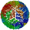
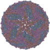
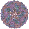
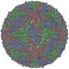
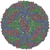
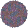
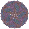
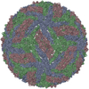
- PDB-5iz7: Cryo-EM structure of thermally stable Zika virus strain H/PF/2013 -
+ Open data

- Basic information
| Entry | Database: PDB / ID: 5iz7 | ||||||||||||
|---|---|---|---|---|---|---|---|---|---|---|---|---|---|
| Title | Cryo-EM structure of thermally stable Zika virus strain H/PF/2013 | ||||||||||||
 Components Components |
| ||||||||||||
 Keywords Keywords |  VIRUS / viral protein / flavivirus / glycoprotein / zika virus VIRUS / viral protein / flavivirus / glycoprotein / zika virus | ||||||||||||
| Function / homology |  Function and homology information Function and homology informationsymbiont-mediated suppression of host JAK-STAT cascade via inhibition of host TYK2 activity / flavivirin / symbiont-mediated suppression of host JAK-STAT cascade via inhibition of STAT2 activity / symbiont-mediated suppression of host JAK-STAT cascade via inhibition of STAT1 activity / ribonucleoside triphosphate phosphatase activity / negative regulation of innate immune response / viral capsid / nucleoside-triphosphate phosphatase / double-stranded RNA binding / 4 iron, 4 sulfur cluster binding ...symbiont-mediated suppression of host JAK-STAT cascade via inhibition of host TYK2 activity / flavivirin / symbiont-mediated suppression of host JAK-STAT cascade via inhibition of STAT2 activity / symbiont-mediated suppression of host JAK-STAT cascade via inhibition of STAT1 activity / ribonucleoside triphosphate phosphatase activity / negative regulation of innate immune response / viral capsid / nucleoside-triphosphate phosphatase / double-stranded RNA binding / 4 iron, 4 sulfur cluster binding / mRNA (guanine-N7)-methyltransferase / methyltransferase cap1 / clathrin-dependent endocytosis of virus by host cell / mRNA (nucleoside-2'-O-)-methyltransferase activity / mRNA 5'-cap (guanine-N7-)-methyltransferase activity / RNA helicase activity / host cell endoplasmic reticulum membrane / host cell perinuclear region of cytoplasm / protein dimerization activity / molecular adaptor activity / RNA helicase / induction by virus of host autophagy / RNA-directed RNA polymerase / symbiont entry into host cell / viral RNA genome replication / RNA-dependent RNA polymerase activity / serine-type endopeptidase activity / fusion of virus membrane with host endosome membrane / centrosome / viral envelope / lipid binding / symbiont-mediated suppression of host type I interferon-mediated signaling pathway / host cell nucleus / structural molecule activity / virion attachment to host cell / GTP binding / virion membrane / ATP hydrolysis activity / proteolysis / extracellular region / ATP binding / membrane / metal ion bindingSimilarity search - Function | ||||||||||||
| Biological species |   Zika virus Zika virus | ||||||||||||
| Method | ELECTRON MICROSCOPY / single particle reconstruction / cryo EM / Resolution: 3.7 Å | ||||||||||||
 Authors Authors | Kostyuchenko, V.A. / Zhang, S. / Fibriansah, G. / Lok, S.M. | ||||||||||||
| Funding support |  Singapore, 3items Singapore, 3items
| ||||||||||||
 Citation Citation | Journal: Nature / Year: 2016 Title: Structure of the thermally stable Zika virus. Authors: Victor A Kostyuchenko / Elisa X Y Lim / Shuijun Zhang / Guntur Fibriansah / Thiam-Seng Ng / Justin S G Ooi / Jian Shi / Shee-Mei Lok / Abstract: Zika virus (ZIKV), formerly a neglected pathogen, has recently been associated with microcephaly in fetuses, and with Guillian-Barré syndrome in adults. Here we present the 3.7 Å resolution cryo- ...Zika virus (ZIKV), formerly a neglected pathogen, has recently been associated with microcephaly in fetuses, and with Guillian-Barré syndrome in adults. Here we present the 3.7 Å resolution cryo-electron microscopy structure of ZIKV, and show that the overall architecture of the virus is similar to that of other flaviviruses. Sequence and structural comparisons of the ZIKV envelope (E) protein with other flaviviruses show that parts of the E protein closely resemble the neurovirulent West Nile and Japanese encephalitis viruses, while others are similar to dengue virus (DENV). However, the contribution of the E protein to flavivirus pathobiology is currently not understood. The virus particle was observed to be structurally stable even when incubated at 40 °C, in sharp contrast to the less thermally stable DENV. This is also reflected in the infectivity of ZIKV compared to DENV serotypes 2 and 4 (DENV2 and DENV4) at different temperatures. The cryo-electron microscopy structure shows a virus with a more compact surface. This structural stability of the virus may help it to survive in the harsh conditions of semen, saliva and urine. Antibodies or drugs that destabilize the structure may help to reduce the disease outcome or limit the spread of the virus. | ||||||||||||
| History |
|
- Structure visualization
| Movie |
Movie viewer |
|---|---|
| Structure viewer | Molecule: MolmilJmol/JSmol |
- Downloads & links
-Download
| PDBx/mmCIF format | 5iz7.cif.gz | 333.5 KB | Display | PDBx/mmCIF format |
|---|---|---|---|---|
| PDB format | pdb5iz7.ent.gz | 268.9 KB | Display | PDB format |
| PDBx/mmJSON format | 5iz7.json.gz | Tree view | PDBx/mmJSON format | |
| Others |  Other downloads Other downloads |
-Validation report
| Arichive directory | https://data.pdbj.org/pub/pdb/validation_reports/iz/5iz7ftp://data.pdbj.org/pub/pdb/validation_reports/iz/5iz7 | HTTPS FTP |
|---|
-Related structure data
| Related structure data | 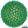 8139MC M: map data used to model this data C: citing same article ( |
|---|---|
| Similar structure data |

-Links
 PDBj
PDBj


- Assembly
| Deposited unit | 
|
|---|---|
| 1 | x 60
|
| 2 |
|
| 3 | x 5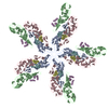
|
| 4 | x 6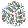
|
| 5 | 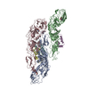
|
| Symmetry | Point symmetry: (Schoenflies symbol: I (icosahedral)) |
-Components
| #1: Protein | Structure Mass: 54444.051 Da / Num. of mol.: 3 / Source method: isolated from a natural source / Details: glycosylated at N154 / Source: (natural) Zika virus / Cell line: C6/36 / Strain: H/PF/2013 / References: UniProt: A0A024B7W1#2: Protein | StructureMass: 8496.883 Da / Num. of mol.: 3 / Source method: isolated from a natural source / Source: (natural) Zika virus / Cell line: C6/36 / Strain: H/PF/2013 / References: UniProt: A0A0U4DG08, UniProt: A0A024B7W1*PLUS#3: Sugar | N-Acetylglucosamine  Type: D-saccharide, beta linking / Mass: 221.208 Da / Num. of mol.: 3 Type: D-saccharide, beta linking / Mass: 221.208 Da / Num. of mol.: 3Source method: isolated from a genetically manipulated source Formula: C8H15NO6 |
|---|
-Experimental details
-Experiment
| Experiment | Method: ELECTRON MICROSCOPY |
|---|---|
| EM experiment | Aggregation state: PARTICLE / 3D reconstruction method: single particle reconstruction |
- Sample preparation
| Component | Name: Zika virus / Type: VIRUS / Entity ID: #1-#2 / Source: NATURAL | ||||||||||||||||||||
|---|---|---|---|---|---|---|---|---|---|---|---|---|---|---|---|---|---|---|---|---|---|
| Molecular weight | Value: 10 MDa / Experimental value: NO | ||||||||||||||||||||
| Source (natural) | Organism: Zika virus / Strain: H/PF/2013 | ||||||||||||||||||||
| Details of virus | Empty: NO / Enveloped: YES / Isolate: STRAIN / Type: VIRION | ||||||||||||||||||||
| Natural host | Organism: Homo sapiens | ||||||||||||||||||||
| Virus shell | Name: glycoprotein shell / Diameter: 480 nm / Triangulation number (T number): 1 | ||||||||||||||||||||
| Buffer solution | pH: 8 | ||||||||||||||||||||
| Buffer component |
| ||||||||||||||||||||
| Specimen | Embedding applied: NO / Shadowing applied: NO / Staining applied: NO / Vitrification applied: YES Details: purified with PEG precipitation, sucrose cushion and potassium tartrate gradient | ||||||||||||||||||||
| Specimen support | Grid material: COPPER / Grid mesh size: 400 divisions/in. / Grid type: Quantifoil | ||||||||||||||||||||
| Vitrification | Instrument: FEI VITROBOT MARK IV / Cryogen name: ETHANE / Humidity: 100 % / Chamber temperature: 298 K / Details: blot for 1s |
- Electron microscopy imaging
| Experimental equipment |  Model: Titan Krios / Image courtesy: FEI Company |
|---|---|
| Microscopy | Model: FEI TITAN KRIOS |
| Electron gun | Electron source: FIELD EMISSION GUN / Accelerating voltage: 300 kV / Illumination mode: FLOOD BEAM |
| Electron lens | Mode: BRIGHT FIELDBright-field microscopy / Cs: 2.7 mm |
| Image recording | Average exposure time: 1.8 sec. / Electron dose: 50 e/Å2 / Detector mode: OTHER / Film or detector model: FEI FALCON II (4k x 4k) / Num. of grids imaged: 1 / Num. of real images: 3086 |
| Image scans | Sampling size: 14 µm / Width: 4096 / Height: 4096 / Movie frames/image: 30 / Used frames/image: 2-11 |
- Processing
| EM software |
| ||||||||||||||||||||||||||||||||||||||||
|---|---|---|---|---|---|---|---|---|---|---|---|---|---|---|---|---|---|---|---|---|---|---|---|---|---|---|---|---|---|---|---|---|---|---|---|---|---|---|---|---|---|
| Image processing | Details: images collected in movie mode, 30 frames per 1.8s exposure; frames aligned with MotionCorr, frames 2-11 averaged to produce images for further processing | ||||||||||||||||||||||||||||||||||||||||
| CTF correction | Details: correction for astigmatism included / Type: PHASE FLIPPING AND AMPLITUDE CORRECTION | ||||||||||||||||||||||||||||||||||||||||
| Particle selection | Num. of particles selected: 11600 / Details: manually picked | ||||||||||||||||||||||||||||||||||||||||
| Symmetry | Point symmetry: I (icosahedral) | ||||||||||||||||||||||||||||||||||||||||
| 3D reconstruction | Resolution: 3.7 Å / Resolution method: FSC 0.143 CUT-OFF / Num. of particles: 7180 / Algorithm: FOURIER SPACE / Num. of class averages: 7180 / Symmetry type: POINT | ||||||||||||||||||||||||||||||||||||||||
| Atomic model building | Protocol: BACKBONE TRACE / Space: RECIPROCAL / Target criteria: residual Details: Only icosahedral symmetry imposed. Rwork 0.323 Rtest 0.324 | ||||||||||||||||||||||||||||||||||||||||
| Atomic model building |
|