 Movie
Movie Controller


+ Open data

- Basic information
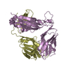
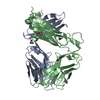
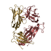
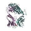
| Entry | Database: PDB / ID: 5iw1 | ||||||
|---|---|---|---|---|---|---|---|
| Title | Crystal Structure of B4.2.3 T-Cell Receptor | ||||||
 Components Components |
| ||||||
 Keywords Keywords |  IMMUNE SYSTEM / T-cell Receptor / TCR / B4.2.3 / B423 / MHC Class I / MOLECULAR RECOGNITION / HUMAN IMMUNODEFICIENCY VIRUS IMMUNE SYSTEM / T-cell Receptor / TCR / B4.2.3 / B423 / MHC Class I / MOLECULAR RECOGNITION / HUMAN IMMUNODEFICIENCY VIRUS | ||||||
| Function / homology | Immunoglobulins / Immunoglobulin-like / Sandwich / Mainly Beta Function and homology information Function and homology information | ||||||
| Biological species |  Mus musculus (house mouse) Mus musculus (house mouse) | ||||||
| Method | X-RAY DIFFRACTION / SYNCHROTRON / MOLECULAR REPLACEMENT / Resolution: 3.001 Å | ||||||
 Authors Authors | Natarajan, K. / Jiang, J. / Margulies, D. | ||||||
 Citation Citation | Journal: Nat Commun / Year: 2017 Title: An allosteric site in the T-cell receptor C beta domain plays a critical signalling role. Authors: Natarajan, K. / McShan, A.C. / Jiang, J. / Kumirov, V.K. / Wang, R. / Zhao, H. / Schuck, P. / Tilahun, M.E. / Boyd, L.F. / Ying, J. / Bax, A. / Margulies, D.H. / Sgourakis, N.G. | ||||||
| History |
|
- Structure visualization
| Structure viewer | Molecule: MolmilJmol/JSmol |
|---|
- Downloads & links
-Download
| PDBx/mmCIF format | 5iw1.cif.gz | 257.4 KB | Display | PDBx/mmCIF format |
|---|---|---|---|---|
| PDB format | pdb5iw1.ent.gz | 208.4 KB | Display | PDB format |
| PDBx/mmJSON format | 5iw1.json.gz | Tree view | PDBx/mmJSON format | |
| Others |  Other downloads Other downloads |
-Validation report
| Arichive directory | https://data.pdbj.org/pub/pdb/validation_reports/iw/5iw1ftp://data.pdbj.org/pub/pdb/validation_reports/iw/5iw1 | HTTPS FTP |
|---|
-Related structure data
| Related structure data |  5ivxSC S: Starting model for refinement C: citing same article ( |
|---|---|
| Similar structure data |


-Links
 PDBj
PDBj

- Assembly
| Deposited unit | 
| ||||||||
|---|---|---|---|---|---|---|---|---|---|
| 1 | 
| ||||||||
| 2 | 
| ||||||||
| 3 | 
| ||||||||
| Unit cell |
|
-Components
| #1: Protein | Mass: 21658.111 Da / Num. of mol.: 3 / Mutation: T163C Source method: isolated from a genetically manipulated source Source: (gene. exp.) Mus musculus (house mouse) / Strain: BALB/C / Cell: T LYMPHOCYTE / Cell line: B4.2.3 T CELL HYBRIDOMA / Gene: TCRAV2S6J38 / Plasmid: PET3A / Production host:  Escherichia coli (E. coli) / Strain (production host): BL21(DE3) Escherichia coli (E. coli) / Strain (production host): BL21(DE3)#2: Protein | Mass: 26909.424 Da / Num. of mol.: 3 / Mutation: S167C, C181A Source method: isolated from a genetically manipulated source Source: (gene. exp.) Mus musculus (house mouse) / Strain: BALB/C / Cell: T LYMPHOCYTE / Cell line: B4.2.3 T CELL HYBRIDOMA / Gene: TCRAV2S6J38 / Plasmid: PET3A / Production host: Escherichia coli (E. coli) / Strain (production host): BL21(DE3) |
|---|
-Experimental details
-Experiment
| Experiment | Method: X-RAY DIFFRACTION / Number of used crystals: 1 |
|---|
- Sample preparation
| Crystal | Density Matthews: 3.18 Å3/Da / Density % sol: 61.3 % |
|---|---|
| Crystal grow | Temperature: 291 K / Method: vapor diffusion, hanging drop / pH: 8.8 / Details: 16% PEG 4000, 0.1M TRIS, 0.2M Magnesium Chloride |
-Data collection
| Diffraction | Mean temperature: 100 K |
|---|---|
| Diffraction source | Source: SYNCHROTRON / Site: NSLS  / Beamline: X29A / Wavelength: 1.0809 Å / Beamline: X29A / Wavelength: 1.0809 Å |
| Detector | Type: ADSC QUANTUM 315 / Detector: CCD / Date: Nov 7, 2006 |
| Radiation | Monochromator: SI III / Protocol: SINGLE WAVELENGTH / Monochromatic (M) / Laue (L): M / Scattering type: x-ray |
| Radiation wavelength | Wavelength: 1.0809 Å / Relative weight: 1 |
| Reflection | Resolution: 3→48.1 Å / Num. obs: 32680 / % possible obs: 94.4 % / Redundancy: 5.4 % / Biso Wilson estimate: 70.3 Å2 / Rmerge(I) obs: 0.131 / Net I/σ(I): 12.6 |
| Reflection shell | Resolution: 3→3.1 Å / Redundancy: 3.8 % / Rmerge(I) obs: 0.482 / Mean I/σ(I) obs: 1.8 / % possible all: 65.3 |
- Processing
| Software |
| |||||||||||||||||||||||||||||||||||||||||||||||||||||||||||||||||||||||||||||||||||||||||||||||||||||||||
|---|---|---|---|---|---|---|---|---|---|---|---|---|---|---|---|---|---|---|---|---|---|---|---|---|---|---|---|---|---|---|---|---|---|---|---|---|---|---|---|---|---|---|---|---|---|---|---|---|---|---|---|---|---|---|---|---|---|---|---|---|---|---|---|---|---|---|---|---|---|---|---|---|---|---|---|---|---|---|---|---|---|---|---|---|---|---|---|---|---|---|---|---|---|---|---|---|---|---|---|---|---|---|---|---|---|---|
| Refinement | Method to determine structure: MOLECULAR REPLACEMENT Starting model: 5IVX Resolution: 3.001→48.056 Å / Cross valid method: THROUGHOUT / σ(F): 1.98 / Phase error: 31.01
| |||||||||||||||||||||||||||||||||||||||||||||||||||||||||||||||||||||||||||||||||||||||||||||||||||||||||
| Solvent computation | Shrinkage radii: 0.9 Å / VDW probe radii: 1.11 Å | |||||||||||||||||||||||||||||||||||||||||||||||||||||||||||||||||||||||||||||||||||||||||||||||||||||||||
| Displacement parameters | Biso mean: 91 Å2 | |||||||||||||||||||||||||||||||||||||||||||||||||||||||||||||||||||||||||||||||||||||||||||||||||||||||||
| Refinement step | Cycle: LAST / Resolution: 3.001→48.056 Å
| |||||||||||||||||||||||||||||||||||||||||||||||||||||||||||||||||||||||||||||||||||||||||||||||||||||||||
| Refine LS restraints |
| |||||||||||||||||||||||||||||||||||||||||||||||||||||||||||||||||||||||||||||||||||||||||||||||||||||||||
| LS refinement shell |
|
