 Movie
Movie Controller

[English] 日本語
 Yorodumi
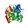
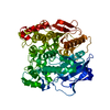
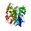
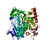
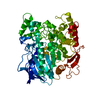
- PDB-5ivk: The alpha-esterase-7 carboxylesterase, E3, from the blowfly Lucil... -
+ Open data

- Basic information
| Entry | Database: PDB / ID: 5ivk | ||||||
|---|---|---|---|---|---|---|---|
| Title | The alpha-esterase-7 carboxylesterase, E3, from the blowfly Lucilia cuprina: phosphorylated-enzyme ensemble refinement | ||||||
 Components Components | Carboxylic ester hydrolase Carboxylesterase Carboxylesterase | ||||||
 Keywords Keywords | HYDROLASE / carboxylesterase / organophosphate / protein dynamics / acetylcholinesterase | ||||||
| Function / homology |  Function and homology informationHydrolases; Acting on ester bonds; Carboxylic-ester hydrolases / carboxylic ester hydrolase activity Function and homology informationHydrolases; Acting on ester bonds; Carboxylic-ester hydrolases / carboxylic ester hydrolase activitySimilarity search - Function | ||||||
| Biological species |  Lucilia cuprina (Australian sheep blowfly) Lucilia cuprina (Australian sheep blowfly) | ||||||
| Method | X-RAY DIFFRACTION / SYNCHROTRON / Resolution: 1.53 Å | ||||||
 Authors Authors | Correy, G.J. / Jackson, C.J. | ||||||
 Citation Citation | Journal: Structure / Year: 2016 Title: Mapping the Accessible Conformational Landscape of an Insect Carboxylesterase Using Conformational Ensemble Analysis and Kinetic Crystallography. Authors: Correy, G.J. / Carr, P.D. / Meirelles, T. / Mabbitt, P.D. / Fraser, N.J. / Weik, M. / Jackson, C.J. | ||||||
| History |
|
- Structure visualization
| Structure viewer | Molecule: MolmilJmol/JSmol |
|---|
- Downloads & links
-Download
| PDBx/mmCIF format | 5ivk.cif.gz | 17 MB | Display | PDBx/mmCIF format |
|---|---|---|---|---|
| PDB format | pdb5ivk.ent.gz | 15 MB | Display | PDB format |
| PDBx/mmJSON format | 5ivk.json.gz | Tree view | PDBx/mmJSON format | |
| Others |  Other downloads Other downloads |
-Validation report
| Arichive directory | https://data.pdbj.org/pub/pdb/validation_reports/iv/5ivkftp://data.pdbj.org/pub/pdb/validation_reports/iv/5ivk | HTTPS FTP |
|---|
-Related structure data
| Related structure data |  4qwmC  4ubiC  4ubjC  4ubkC  4ublC  4ubmC  4ubnC  4uboC  4w1pC  4w1qC  4w1rC  4w1sC  5ch3C  5ch5C  5ivdC  5ivhC  5iviC C: citing same article ( |
|---|---|
| Similar structure data |


-Links
 PDBj
PDBj
- Assembly
| Deposited unit | 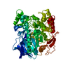
| |||||||||||||||||||||||||||||||||||||||||||||||||||||||||||||||||||||||||||||||||||||||||||||||||||||||||||||||||||||||||||||||||||||||
|---|---|---|---|---|---|---|---|---|---|---|---|---|---|---|---|---|---|---|---|---|---|---|---|---|---|---|---|---|---|---|---|---|---|---|---|---|---|---|---|---|---|---|---|---|---|---|---|---|---|---|---|---|---|---|---|---|---|---|---|---|---|---|---|---|---|---|---|---|---|---|---|---|---|---|---|---|---|---|---|---|---|---|---|---|---|---|---|---|---|---|---|---|---|---|---|---|---|---|---|---|---|---|---|---|---|---|---|---|---|---|---|---|---|---|---|---|---|---|---|---|---|---|---|---|---|---|---|---|---|---|---|---|---|---|---|---|
| 1 |
| |||||||||||||||||||||||||||||||||||||||||||||||||||||||||||||||||||||||||||||||||||||||||||||||||||||||||||||||||||||||||||||||||||||||
| Unit cell |
| |||||||||||||||||||||||||||||||||||||||||||||||||||||||||||||||||||||||||||||||||||||||||||||||||||||||||||||||||||||||||||||||||||||||
| Number of models | 43 | |||||||||||||||||||||||||||||||||||||||||||||||||||||||||||||||||||||||||||||||||||||||||||||||||||||||||||||||||||||||||||||||||||||||
| Components on special symmetry positions |
|
-Components
| #1: Protein | Carboxylesterase Mass: 66432.844 Da / Num. of mol.: 1 Source method: isolated from a genetically manipulated source Source: (gene. exp.) Lucilia cuprina (Australian sheep blowfly)Gene: LcaE7 / Production host:  Escherichia coli (E. coli) Escherichia coli (E. coli)References: UniProt: Q25252, Hydrolases; Acting on ester bonds; Carboxylic-ester hydrolases |
|---|---|
| #2: Chemical | ChemComp-DPF /   Mass: 154.102 Da / Num. of mol.: 1 Mass: 154.102 Da / Num. of mol.: 1Source method: isolated from a genetically manipulated source Formula: C4H11O4P |
| #3: Water | ChemComp-HOH / Water Mass: 18.015 Da / Num. of mol.: 399 / Source method: isolated from a natural source / Formula: H2O Mass: 18.015 Da / Num. of mol.: 399 / Source method: isolated from a natural source / Formula: H2O |
-Experimental details
-Experiment
| Experiment | Method: X-RAY DIFFRACTION / Number of used crystals: 1 |
|---|
- Sample preparation
| Crystal | Density Matthews: 2.26 Å3/Da / Density % sol: 45.62 % |
|---|---|
| Crystal grow | Temperature: 298 K / Method: vapor diffusion, hanging drop / Details: 100mM sodium acetate pH 4.5, PEG 2K MME |
-Data collection
| Diffraction | Mean temperature: 100 K |
|---|---|
| Diffraction source | Source: SYNCHROTRON / Site: ESRF  / Beamline: ID23-2 / Wavelength: 0.8266 Å / Beamline: ID23-2 / Wavelength: 0.8266 Å |
| Detector | Type: ADSC QUANTUM 315r / Detector: CCD / Date: May 12, 2010 |
| Radiation | Protocol: SINGLE WAVELENGTH / Monochromatic (M) / Laue (L): M / Scattering type: x-ray |
| Radiation wavelength | Wavelength: 0.8266 Å / Relative weight: 1 |
| Reflection | Resolution: 1.53→42 Å / Num. obs: 89882 / % possible obs: 100 % / Redundancy: 12.7 % / Net I/σ(I): 11.6 |
| Reflection shell | Resolution: 1.53→1.56 Å / Redundancy: 12 % / Rmerge(I) obs: 1.79 / Mean I/σ(I) obs: 1.4 / % possible all: 100 |
- Processing
| Software |
| |||||||||||||||||||||||||||||||||||||||||||||||||||||||||||||||||||||||||||||||||||||||||||||||||||||||||||||||||||||||||||||||||||||||||||||||||||||||||||||||||||||||||||||||||||||||||||||||||||||||||||||||||||||||||
|---|---|---|---|---|---|---|---|---|---|---|---|---|---|---|---|---|---|---|---|---|---|---|---|---|---|---|---|---|---|---|---|---|---|---|---|---|---|---|---|---|---|---|---|---|---|---|---|---|---|---|---|---|---|---|---|---|---|---|---|---|---|---|---|---|---|---|---|---|---|---|---|---|---|---|---|---|---|---|---|---|---|---|---|---|---|---|---|---|---|---|---|---|---|---|---|---|---|---|---|---|---|---|---|---|---|---|---|---|---|---|---|---|---|---|---|---|---|---|---|---|---|---|---|---|---|---|---|---|---|---|---|---|---|---|---|---|---|---|---|---|---|---|---|---|---|---|---|---|---|---|---|---|---|---|---|---|---|---|---|---|---|---|---|---|---|---|---|---|---|---|---|---|---|---|---|---|---|---|---|---|---|---|---|---|---|---|---|---|---|---|---|---|---|---|---|---|---|---|---|---|---|---|---|---|---|---|---|---|---|---|---|---|---|---|---|---|---|---|
| Refinement | Resolution: 1.53→42.01 Å / SU ML: 0.15 / Cross valid method: FREE R-VALUE / σ(F): 1.34 / Phase error: 21.81
| |||||||||||||||||||||||||||||||||||||||||||||||||||||||||||||||||||||||||||||||||||||||||||||||||||||||||||||||||||||||||||||||||||||||||||||||||||||||||||||||||||||||||||||||||||||||||||||||||||||||||||||||||||||||||
| Solvent computation | Shrinkage radii: 0.8 Å / VDW probe radii: 1 Å / Solvent model: FLAT BULK SOLVENT MODEL | |||||||||||||||||||||||||||||||||||||||||||||||||||||||||||||||||||||||||||||||||||||||||||||||||||||||||||||||||||||||||||||||||||||||||||||||||||||||||||||||||||||||||||||||||||||||||||||||||||||||||||||||||||||||||
| Refinement step | Cycle: LAST / Resolution: 1.53→42.01 Å
| |||||||||||||||||||||||||||||||||||||||||||||||||||||||||||||||||||||||||||||||||||||||||||||||||||||||||||||||||||||||||||||||||||||||||||||||||||||||||||||||||||||||||||||||||||||||||||||||||||||||||||||||||||||||||
| Refine LS restraints |
| |||||||||||||||||||||||||||||||||||||||||||||||||||||||||||||||||||||||||||||||||||||||||||||||||||||||||||||||||||||||||||||||||||||||||||||||||||||||||||||||||||||||||||||||||||||||||||||||||||||||||||||||||||||||||
| LS refinement shell |
|
