 Movie
Movie Controller

[English] 日本語
 Yorodumi
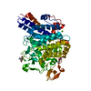
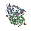
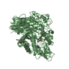
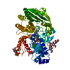
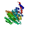
- PDB-5iv4: Crystal structure of the human soluble adenylyl cyclase in comple... -
+ Open data

- Basic information
| Entry | Database: PDB / ID: 5iv4 | ||||||
|---|---|---|---|---|---|---|---|
| Title | Crystal structure of the human soluble adenylyl cyclase in complex with the allosteric inhibitor LRE1 | ||||||
 Components Components | Adenylate cyclase type 10 | ||||||
 Keywords Keywords |  LYASE / human soluble Adenylyl Cyclase complex LRE1 inhibitor LYASE / human soluble Adenylyl Cyclase complex LRE1 inhibitor | ||||||
| Function / homology |  Function and homology information Function and homology informationnegative regulation of cardiac muscle cell contraction / astrocyte end-foot / mitochondrial ATP transmembrane transport / bicarbonate binding / epithelial cilium movement involved in extracellular fluid movement / neuron projection retraction / positive regulation of glycogen catabolic process / : / central region of growth cone / regulation of mitophagy ...negative regulation of cardiac muscle cell contraction / astrocyte end-foot / mitochondrial ATP transmembrane transport / bicarbonate binding / epithelial cilium movement involved in extracellular fluid movement / neuron projection retraction / positive regulation of glycogen catabolic process / : / central region of growth cone / regulation of mitophagy / glucose catabolic process / regulation of membrane repolarization / adenylate cyclase / basal part of cell / positive regulation of oxidative stress-induced neuron intrinsic apoptotic signaling pathway / cAMP biosynthetic process / positive regulation of ossification / adenylate cyclase activity / positive regulation of protein targeting to mitochondrion / positive regulation of reactive oxygen species biosynthetic process / neuron projection extension / positive regulation of vascular associated smooth muscle cell apoptotic process / positive regulation of mitochondrial depolarization / positive regulation of ATP biosynthetic process / positive regulation of cardiac muscle hypertrophy / positive regulation of cardiac muscle cell apoptotic process / negative regulation of mitochondrial membrane potential / spermatid development / positive regulation of axon extension / Hedgehog 'off' state / negative regulation of reactive oxygen species biosynthetic process / neuron projection maintenance / positive regulation of peptidyl-threonine phosphorylation / cilium / manganese ion binding / ATPase binding / cytoskeleton / intracellular signal transduction / apical plasma membrane / neuronal cell body / dendrite / perinuclear region of cytoplasm / magnesium ion binding / mitochondrion / extracellular region / ATP binding / nucleus / cytosol / cytoplasmSimilarity search - Function | ||||||
| Biological species |  Homo sapiens (human) Homo sapiens (human) | ||||||
| Method | X-RAY DIFFRACTION / SYNCHROTRON / Resolution: 1.79 Å | ||||||
 Authors Authors | Kleinboelting, S. / Steegborn, C. | ||||||
| Funding support |  Germany, 1items Germany, 1items
| ||||||
 Citation Citation | Journal: Nat.Chem.Biol. / Year: 2016 Title: Discovery of LRE1 as a specific and allosteric inhibitor of soluble adenylyl cyclase. Authors: Ramos-Espiritu, L. / Kleinboelting, S. / Navarrete, F.A. / Alvau, A. / Visconti, P.E. / Valsecchi, F. / Starkov, A. / Manfredi, G. / Buck, H. / Adura, C. / Zippin, J.H. / van den Heuvel, J. ...Authors: Ramos-Espiritu, L. / Kleinboelting, S. / Navarrete, F.A. / Alvau, A. / Visconti, P.E. / Valsecchi, F. / Starkov, A. / Manfredi, G. / Buck, H. / Adura, C. / Zippin, J.H. / van den Heuvel, J. / Glickman, J.F. / Steegborn, C. / Levin, L.R. / Buck, J. | ||||||
| History |
|
- Structure visualization
| Structure viewer | Molecule: MolmilJmol/JSmol |
|---|
- Downloads & links
-Download
| PDBx/mmCIF format | 5iv4.cif.gz | 116.3 KB | Display | PDBx/mmCIF format |
|---|---|---|---|---|
| PDB format | pdb5iv4.ent.gz | 91 KB | Display | PDB format |
| PDBx/mmJSON format | 5iv4.json.gz | Tree view | PDBx/mmJSON format | |
| Others |  Other downloads Other downloads |
-Validation report
| Arichive directory | https://data.pdbj.org/pub/pdb/validation_reports/iv/5iv4ftp://data.pdbj.org/pub/pdb/validation_reports/iv/5iv4 | HTTPS FTP |
|---|
-Related structure data


-Links
 PDBj
PDBj


- Assembly
| Deposited unit | 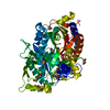
| ||||||||
|---|---|---|---|---|---|---|---|---|---|
| 1 |
| ||||||||
| Unit cell |
|
-Components
-Protein , 1 types, 1 molecules A
| #1: Protein | Mass: 54269.664 Da / Num. of mol.: 1 Source method: isolated from a genetically manipulated source Details: Cys255 modified by BME which is in the purification buffer Source: (gene. exp.) Homo sapiens (human) / Gene: ADCY10, SAC / Production host:  Trichoplusia ni (cabbage looper) / References: UniProt: Q96PN6, adenylate cyclase Trichoplusia ni (cabbage looper) / References: UniProt: Q96PN6, adenylate cyclase |
|---|
-Non-polymers , 8 types, 373 molecules 


| #2: Chemical | ChemComp-LRI /  Mass: 280.776 Da / Num. of mol.: 1 / Source method: obtained synthetically / Formula: C12H13ClN4S Mass: 280.776 Da / Num. of mol.: 1 / Source method: obtained synthetically / Formula: C12H13ClN4S | ||||||||||||
|---|---|---|---|---|---|---|---|---|---|---|---|---|---|
| #3: Chemical | Acetate Mass: 59.044 Da / Num. of mol.: 2 / Source method: obtained synthetically / Formula: C2H3O2 Mass: 59.044 Da / Num. of mol.: 2 / Source method: obtained synthetically / Formula: C2H3O2#4: Chemical | ChemComp-EDO / Ethylene glycol Mass: 62.068 Da / Num. of mol.: 5 / Source method: obtained synthetically / Formula: C2H6O2 Mass: 62.068 Da / Num. of mol.: 5 / Source method: obtained synthetically / Formula: C2H6O2#5: Chemical | ChemComp-DMS / Dimethyl sulfoxide Mass: 78.133 Da / Num. of mol.: 4 / Source method: obtained synthetically / Formula: C2H6OS / Comment: DMSO, precipitant*YM Mass: 78.133 Da / Num. of mol.: 4 / Source method: obtained synthetically / Formula: C2H6OS / Comment: DMSO, precipitant*YM#6: Chemical | ChemComp-CL / | Chloride Mass: 35.453 Da / Num. of mol.: 1 / Source method: obtained synthetically / Formula: Cl Mass: 35.453 Da / Num. of mol.: 1 / Source method: obtained synthetically / Formula: Cl#7: Chemical | ChemComp-PEG / | Diethylene glycol Mass: 106.120 Da / Num. of mol.: 1 / Source method: obtained synthetically / Formula: C4H10O3 Mass: 106.120 Da / Num. of mol.: 1 / Source method: obtained synthetically / Formula: C4H10O3#8: Chemical | ChemComp-GOL / | Glycerol Mass: 92.094 Da / Num. of mol.: 1 / Source method: obtained synthetically / Formula: C3H8O3 Mass: 92.094 Da / Num. of mol.: 1 / Source method: obtained synthetically / Formula: C3H8O3#9: Water | ChemComp-HOH / | WaterMass: 18.015 Da / Num. of mol.: 358 / Source method: isolated from a natural source / Formula: H2O |
-Experimental details
-Experiment
| Experiment | Method: X-RAY DIFFRACTION / Number of used crystals: 1 |
|---|
- Sample preparation
| Crystal | Density Matthews: 2.3 Å3/Da / Density % sol: 47 % |
|---|---|
| Crystal grow | Temperature: 277.15 K / Method: vapor diffusion, hanging drop / pH: 6.5 Details: 100 mM sodium acetate pH 4.8 200 mM tri-sodium-citrate 15% (w/v) PEG 4000 10% (v/v) glycerol |
-Data collection
| Diffraction | Mean temperature: 100 K |
|---|---|
| Diffraction source | Source: SYNCHROTRON / Site: BESSY / Beamline: 14.1 / Wavelength: 0.918007 Å |
| Detector | Type: DECTRIS PILATUS 6M / Detector: PIXEL / Date: Feb 4, 2015 |
| Radiation | Protocol: SINGLE WAVELENGTH / Monochromatic (M) / Laue (L): M / Scattering type: x-ray |
| Radiation wavelength | Wavelength: 0.918007 Å / Relative weight: 1 |
| Reflection | Resolution: 1.79→44.42 Å / Num. obs: 599809 / % possible obs: 99.9 % / Redundancy: 11.4 % / Rmerge(I) obs: 0.115 / Net I/σ(I): 16.3 |
| Reflection shell | Resolution: 1.79→1.9 Å / Redundancy: 11.2 % / Rmerge(I) obs: 1.192 / Mean I/σ(I) obs: 2.07 / % possible all: 99.5 |
- Processing
| Software |
| ||||||||||||||||||||||||||||||||||||||||||||||||||||||||||||||||||||||||||||||||||||||||||||||||||||||||||||||||||||||||||||||||||||||||||||||||||||||||||||||||||||||||||||||||||||||
|---|---|---|---|---|---|---|---|---|---|---|---|---|---|---|---|---|---|---|---|---|---|---|---|---|---|---|---|---|---|---|---|---|---|---|---|---|---|---|---|---|---|---|---|---|---|---|---|---|---|---|---|---|---|---|---|---|---|---|---|---|---|---|---|---|---|---|---|---|---|---|---|---|---|---|---|---|---|---|---|---|---|---|---|---|---|---|---|---|---|---|---|---|---|---|---|---|---|---|---|---|---|---|---|---|---|---|---|---|---|---|---|---|---|---|---|---|---|---|---|---|---|---|---|---|---|---|---|---|---|---|---|---|---|---|---|---|---|---|---|---|---|---|---|---|---|---|---|---|---|---|---|---|---|---|---|---|---|---|---|---|---|---|---|---|---|---|---|---|---|---|---|---|---|---|---|---|---|---|---|---|---|---|---|
| Refinement | Resolution: 1.79→44.42 Å / Cor.coef. Fo:Fc: 0.971 / Cor.coef. Fo:Fc free: 0.953 / SU B: 2.647 / SU ML: 0.08 / Cross valid method: THROUGHOUT / ESU R: 0.103 / ESU R Free: 0.109 / Stereochemistry target values: MAXIMUM LIKELIHOOD / Details: HYDROGENS HAVE BEEN ADDED IN THE RIDING POSITIONS
| ||||||||||||||||||||||||||||||||||||||||||||||||||||||||||||||||||||||||||||||||||||||||||||||||||||||||||||||||||||||||||||||||||||||||||||||||||||||||||||||||||||||||||||||||||||||
| Solvent computation | Ion probe radii: 0.8 Å / Shrinkage radii: 0.8 Å / VDW probe radii: 1.2 Å / Solvent model: MASK | ||||||||||||||||||||||||||||||||||||||||||||||||||||||||||||||||||||||||||||||||||||||||||||||||||||||||||||||||||||||||||||||||||||||||||||||||||||||||||||||||||||||||||||||||||||||
| Displacement parameters | Biso mean: 30.704 Å2
| ||||||||||||||||||||||||||||||||||||||||||||||||||||||||||||||||||||||||||||||||||||||||||||||||||||||||||||||||||||||||||||||||||||||||||||||||||||||||||||||||||||||||||||||||||||||
| Refinement step | Cycle: LAST / Resolution: 1.79→44.42 Å
| ||||||||||||||||||||||||||||||||||||||||||||||||||||||||||||||||||||||||||||||||||||||||||||||||||||||||||||||||||||||||||||||||||||||||||||||||||||||||||||||||||||||||||||||||||||||
| Refine LS restraints |
|