 Movie
Movie Controller

[English] 日本語
 Yorodumi
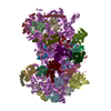
- PDB-5it9: Structure of the yeast Kluyveromyces lactis small ribosomal subun... -
+ Open data

- Basic information
| Entry | Database: PDB / ID: 5it9 | ||||||||||||
|---|---|---|---|---|---|---|---|---|---|---|---|---|---|
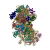
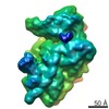
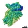
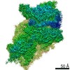
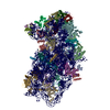
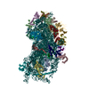
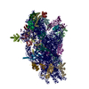
| Title | Structure of the yeast Kluyveromyces lactis small ribosomal subunit in complex with the cricket paralysis virus IRES. | ||||||||||||
 Components Components |
| ||||||||||||
 Keywords Keywords |  RIBOSOME / IRES / small / subunit RIBOSOME / IRES / small / subunit | ||||||||||||
| Function / homology |  Function and homology information Function and homology informationtranslation regulator activity / cytosolic ribosome / ribosomal small subunit assembly / rRNA processing / cytosolic small ribosomal subunit / ribosome binding / small ribosomal subunit / rRNA binding / ribosome / structural constituent of ribosome ...translation regulator activity / cytosolic ribosome / ribosomal small subunit assembly / rRNA processing / cytosolic small ribosomal subunit / ribosome binding / small ribosomal subunit / rRNA binding / ribosome / structural constituent of ribosome / ribonucleoprotein complex / translation / RNA binding / zinc ion binding / metal ion binding / nucleus / cytosol / cytoplasmSimilarity search - Function | ||||||||||||
| Biological species |  Cricket paralysis virus Cricket paralysis virus Kluyveromyces lactis (yeast) Kluyveromyces lactis (yeast) | ||||||||||||
| Method | ELECTRON MICROSCOPY / single particle reconstruction / cryo EM / Resolution: 3.8 Å | ||||||||||||
 Authors Authors | Murray, J. / Savva, C.G. / Shin, B.S. / Dever, T.E. / Ramakrishnan, V. / Fernandez, I.S. | ||||||||||||
| Funding support |  United Kingdom, United Kingdom,  United States, 3items United States, 3items
| ||||||||||||
 Citation Citation | Journal: Elife / Year: 2016 Title: Structural characterization of ribosome recruitment and translocation by type IV IRES. Authors: Jason Murray / Christos G Savva / Byung-Sik Shin / Thomas E Dever / V Ramakrishnan / Israel S Fernández / Abstract: Viral mRNA sequences with a type IV IRES are able to initiate translation without any host initiation factors. Initial recruitment of the small ribosomal subunit as well as two translocation steps ...Viral mRNA sequences with a type IV IRES are able to initiate translation without any host initiation factors. Initial recruitment of the small ribosomal subunit as well as two translocation steps before the first peptidyl transfer are essential for the initiation of translation by these mRNAs. Using electron cryomicroscopy (cryo-EM) we have structurally characterized at high resolution how the Cricket Paralysis Virus Internal Ribosomal Entry Site (CrPV-IRES) binds the small ribosomal subunit (40S) and the translocation intermediate stabilized by elongation factor 2 (eEF2). The CrPV-IRES restricts tvhe otherwise flexible 40S head to a conformation compatible with binding the large ribosomal subunit (60S). Once the 60S is recruited, the binary CrPV-IRES/80S complex oscillates between canonical and rotated states (Fernández et al., 2014; Koh et al., 2014), as seen for pre-translocation complexes with tRNAs. Elongation factor eEF2 with a GTP analog stabilizes the ribosome-IRES complex in a rotated state with an extra ~3 degrees of rotation. Key residues in domain IV of eEF2 interact with pseudoknot I (PKI) of the CrPV-IRES stabilizing it in a conformation reminiscent of a hybrid tRNA state. The structure explains how diphthamide, a eukaryotic and archaeal specific post-translational modification of a histidine residue of eEF2, is involved in translocation. | ||||||||||||
| History |
|
- Structure visualization
| Movie |
Movie viewer |
|---|---|
| Structure viewer | Molecule: MolmilJmol/JSmol |
- Downloads & links
-Download
| PDBx/mmCIF format | 5it9.cif.gz | 1.9 MB | Display | PDBx/mmCIF format |
|---|---|---|---|---|
| PDB format | pdb5it9.ent.gz | 1.6 MB | Display | PDB format |
| PDBx/mmJSON format | 5it9.json.gz | Tree view | PDBx/mmJSON format | |
| Others |  Other downloads Other downloads |
-Validation report
| Arichive directory | https://data.pdbj.org/pub/pdb/validation_reports/it/5it9ftp://data.pdbj.org/pub/pdb/validation_reports/it/5it9 | HTTPS FTP |
|---|
-Related structure data
| Related structure data | 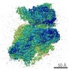 8124MC  8123C  5it7C M: map data used to model this data C: citing same article ( |
|---|---|
| Similar structure data |


-Links
 PDBj
PDBj


- Assembly
| Deposited unit | 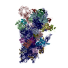
|
|---|---|
| 1 |
|
-Components
+Ribosomal protein ... , 33 types, 33 molecules ABCDEFGHIJKLMNOPQRSTUVWXYZabcd...
-RNA chain , 2 types, 2 molecules 2i
| #34: RNA chain | Mass: 573508.938 Da / Num. of mol.: 1 / Source method: isolated from a natural source / Source: (natural) Kluyveromyces lactis (yeast) |
|---|---|
| #35: RNA chain | Mass: 61462.207 Da / Num. of mol.: 1 Source method: isolated from a genetically manipulated source Source: (gene. exp.) Cricket paralysis virus / Production host:  Escherichia coli (E. coli) / References: GenBank: 8895506 Escherichia coli (E. coli) / References: GenBank: 8895506 |
-Non-polymers , 2 types, 83 molecules 

| #36: Chemical | ChemComp-MG /  Mass: 24.305 Da / Num. of mol.: 80 / Source method: obtained synthetically / Formula: Mg Mass: 24.305 Da / Num. of mol.: 80 / Source method: obtained synthetically / Formula: Mg#37: Chemical |  Mass: 65.409 Da / Num. of mol.: 3 / Source method: obtained synthetically / Formula: Zn Mass: 65.409 Da / Num. of mol.: 3 / Source method: obtained synthetically / Formula: Zn |
|---|
-Experimental details
-Experiment
| Experiment | Method: ELECTRON MICROSCOPY |
|---|---|
| EM experiment | Aggregation state: PARTICLE / 3D reconstruction method: single particle reconstruction |
- Sample preparation
| Component | Name: Kluyveromyces lactis small ribosomal subunit in complex with the Cricket paralysis virus IRES Type: RIBOSOME / Entity ID: #1-#35 / Source: NATURAL | ||||||||||||||||||||
|---|---|---|---|---|---|---|---|---|---|---|---|---|---|---|---|---|---|---|---|---|---|
| Molecular weight | Value: 2 MDa / Experimental value: NO | ||||||||||||||||||||
| Source (natural) | Organism: Kluyveromyces lacti (yeast) | ||||||||||||||||||||
| Buffer solution | pH: 6.5 | ||||||||||||||||||||
| Buffer component |
| ||||||||||||||||||||
| Specimen | Embedding applied: NO / Shadowing applied: NO / Staining applied: NO / Vitrification applied: YES | ||||||||||||||||||||
| Specimen support | Grid material: COPPER / Grid mesh size: 300 divisions/in. / Grid type: Quantifoil | ||||||||||||||||||||
| Vitrification | Instrument: FEI VITROBOT MARK II / Cryogen name: ETHANE / Humidity: 100 % / Chamber temperature: 298 K |
- Electron microscopy imaging
| Experimental equipment |  Model: Titan Krios / Image courtesy: FEI Company |
|---|---|
| Microscopy | Model: FEI TITAN KRIOS |
| Electron gun | Electron source: FIELD EMISSION GUN / Accelerating voltage: 300 kV / Illumination mode: OTHER |
| Electron lens | Mode: BRIGHT FIELDBright-field microscopy |
| Specimen holder | Specimen holder model: FEI TITAN KRIOS AUTOGRID HOLDER |
| Image recording | Electron dose: 25 e/Å2 / Detector mode: INTEGRATING / Film or detector model: FEI FALCON II (4k x 4k) |
- Processing
| Software | Name: REFMAC / Version: 5.8.0124 / Classification: refinement | |||||||||||||||||||||||||||||||||||||||||||||||||||||||||||||||||||||||||||
|---|---|---|---|---|---|---|---|---|---|---|---|---|---|---|---|---|---|---|---|---|---|---|---|---|---|---|---|---|---|---|---|---|---|---|---|---|---|---|---|---|---|---|---|---|---|---|---|---|---|---|---|---|---|---|---|---|---|---|---|---|---|---|---|---|---|---|---|---|---|---|---|---|---|---|---|---|
| EM software |
| |||||||||||||||||||||||||||||||||||||||||||||||||||||||||||||||||||||||||||
| CTF correction | Type: PHASE FLIPPING ONLY | |||||||||||||||||||||||||||||||||||||||||||||||||||||||||||||||||||||||||||
| 3D reconstruction | Resolution: 3.8 Å / Resolution method: FSC 0.143 CUT-OFF / Num. of particles: 54481 / Algorithm: BACK PROJECTION / Symmetry type: POINT | |||||||||||||||||||||||||||||||||||||||||||||||||||||||||||||||||||||||||||
| Atomic model building | Protocol: OTHER / Space: RECIPROCAL | |||||||||||||||||||||||||||||||||||||||||||||||||||||||||||||||||||||||||||
| Refinement | Resolution: 3.8→257.28 Å / Cor.coef. Fo:Fc: 0.848 / SU B: 43.619 / SU ML: 0.614 / σ(F): 0 / ESU R: 0.945 Stereochemistry target values: MAXIMUM LIKELIHOOD WITH PHASES Details: HYDROGENS HAVE BEEN ADDED IN THE RIDING POSITIONS U VALUES : REFINED INDIVIDUALLY
| |||||||||||||||||||||||||||||||||||||||||||||||||||||||||||||||||||||||||||
| Solvent computation | Solvent model: NONE | |||||||||||||||||||||||||||||||||||||||||||||||||||||||||||||||||||||||||||
| Displacement parameters | Biso max: 500 Å2 / Biso mean: 119.994 Å2 / Biso min: 2 Å2
| |||||||||||||||||||||||||||||||||||||||||||||||||||||||||||||||||||||||||||
| Refinement step | Cycle: final / Resolution: 3.8→257.28 Å
| |||||||||||||||||||||||||||||||||||||||||||||||||||||||||||||||||||||||||||
| Refine LS restraints |
| |||||||||||||||||||||||||||||||||||||||||||||||||||||||||||||||||||||||||||
| LS refinement shell | Resolution: 3.8→3.899 Å / Total num. of bins used: 20
|
