 Movie
Movie Controller

[English] 日本語
 Yorodumi
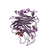
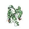
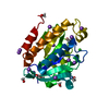
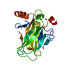
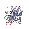
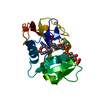
- PDB-5imb: Crystal structure of peptidyl-tRNA hydrolase mutant-N14D from Vib... -
+ Open data

- Basic information
| Entry | Database: PDB / ID: 5imb | ||||||
|---|---|---|---|---|---|---|---|
| Title | Crystal structure of peptidyl-tRNA hydrolase mutant-N14D from Vibrio cholerae | ||||||
 Components Components | Peptidyl-tRNA hydrolase Alternative ribosome-rescue factor B Alternative ribosome-rescue factor B | ||||||
 Keywords Keywords | HYDROLASE / Peptidyl-tRNA hydrolase / N14D mutant / Vibrio cholerae | ||||||
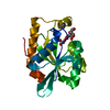
| Function / homology |  Function and homology informationpeptidyl-tRNA hydrolase / aminoacyl-tRNA hydrolase activity / translation / cytoplasm Function and homology informationpeptidyl-tRNA hydrolase / aminoacyl-tRNA hydrolase activity / translation / cytoplasmSimilarity search - Function | ||||||
| Biological species |  Vibrio cholerae O1 biovar El Tor str. N16961 (bacteria) Vibrio cholerae O1 biovar El Tor str. N16961 (bacteria) | ||||||
| Method | X-RAY DIFFRACTION / MOLECULAR REPLACEMENT / Resolution: 2.4 Å | ||||||
 Authors Authors | Shahid, S. / Kabra, A. / Pal, R.K. / Arora, A. | ||||||
 Citation Citation | Journal: RNA / Year: 2017 Title: Unraveling the stereochemical and dynamic aspects of the catalytic site of bacterial peptidyl-tRNA hydrolase. Authors: Kabra, A. / Shahid, S. / Pal, R.K. / Yadav, R. / Pulavarti, S.V. / Jain, A. / Tripathi, S. / Arora, A. | ||||||
| History |
|
- Structure visualization
| Structure viewer | Molecule: MolmilJmol/JSmol |
|---|
- Downloads & links
-Download
| PDBx/mmCIF format | 5imb.cif.gz | 160.6 KB | Display | PDBx/mmCIF format |
|---|---|---|---|---|
| PDB format | pdb5imb.ent.gz | 126.1 KB | Display | PDB format |
| PDBx/mmJSON format | 5imb.json.gz | Tree view | PDBx/mmJSON format | |
| Others |  Other downloads Other downloads |
-Validation report
| Arichive directory | https://data.pdbj.org/pub/pdb/validation_reports/im/5imbftp://data.pdbj.org/pub/pdb/validation_reports/im/5imb | HTTPS FTP |
|---|
-Related structure data
| Related structure data | 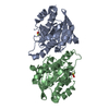 4z86C 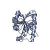 4zxpSC 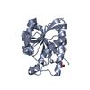 5b6jC  5ikeC S: Starting model for refinement C: citing same article ( |
|---|---|
| Similar structure data |


-Links
 PDBj
PDBj- Assembly
| Deposited unit | 
| ||||||||
|---|---|---|---|---|---|---|---|---|---|
| 1 | 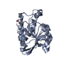
| ||||||||
| 2 | 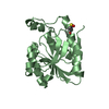
| ||||||||
| Unit cell |
|
-Components
| #1: Protein | Alternative ribosome-rescue factor B / PTH Mass: 21761.152 Da / Num. of mol.: 2 / Mutation: N14D Source method: isolated from a genetically manipulated source Source: (gene. exp.) Vibrio cholerae O1 biovar El Tor str. N16961 (bacteria)Strain: ATCC 39315 / El Tor Inaba N16961 / Gene: pth, VC_2184 / Production host: Escherichia coli (E. coli) / Strain (production host): BL21-Gold(DE3)pLysS AG / References: UniProt: Q9KQ21, peptidyl-tRNA hydrolase#2: Water | ChemComp-HOH / | Water Mass: 18.015 Da / Num. of mol.: 28 / Source method: isolated from a natural source / Formula: H2O Mass: 18.015 Da / Num. of mol.: 28 / Source method: isolated from a natural source / Formula: H2OSequence details | TEV protease site was present cleaved in the purification process | |
|---|
-Experimental details
-Experiment
| Experiment | Method: X-RAY DIFFRACTION / Number of used crystals: 1 |
|---|
- Sample preparation
| Crystal | Density Matthews: 2.31 Å3/Da / Density % sol: 46.78 % |
|---|---|
| Crystal grow | Temperature: 288 K / Method: vapor diffusion, hanging drop Details: 0.1M Sodium citrate, 0.2M Ammonium acetate, 27.5% Polyethylene glycol 4000 |
-Data collection
| Diffraction | Mean temperature: 100 K |
|---|---|
| Diffraction source | Source: ROTATING ANODE / Type: RIGAKU FR-E+ SUPERBRIGHT / Wavelength: 1.5418 Å |
| Detector | Type: RIGAKU RAXIS IV++ / Detector: IMAGE PLATE / Date: Dec 20, 2015 |
| Radiation | Protocol: SINGLE WAVELENGTH / Monochromatic (M) / Laue (L): M / Scattering type: x-ray |
| Radiation wavelength | Wavelength: 1.5418 Å / Relative weight: 1 |
| Reflection | Resolution: 2.4→50 Å / Num. obs: 16047 / % possible obs: 98.3 % / Redundancy: 5.7 % / Net I/σ(I): 28.26 |
| Reflection shell | Resolution: 2.4→2.49 Å |
- Processing
| Software |
| ||||||||||||||||||||||||||||||||||||||||||||||||||||||||||||||||||||||||||||||||||||||||||||||||||||||||||||||||||||||||||||||||||||||||||||||||||||||||||||||||||||||||||||||||||||||
|---|---|---|---|---|---|---|---|---|---|---|---|---|---|---|---|---|---|---|---|---|---|---|---|---|---|---|---|---|---|---|---|---|---|---|---|---|---|---|---|---|---|---|---|---|---|---|---|---|---|---|---|---|---|---|---|---|---|---|---|---|---|---|---|---|---|---|---|---|---|---|---|---|---|---|---|---|---|---|---|---|---|---|---|---|---|---|---|---|---|---|---|---|---|---|---|---|---|---|---|---|---|---|---|---|---|---|---|---|---|---|---|---|---|---|---|---|---|---|---|---|---|---|---|---|---|---|---|---|---|---|---|---|---|---|---|---|---|---|---|---|---|---|---|---|---|---|---|---|---|---|---|---|---|---|---|---|---|---|---|---|---|---|---|---|---|---|---|---|---|---|---|---|---|---|---|---|---|---|---|---|---|---|---|
| Refinement | Method to determine structure: MOLECULAR REPLACEMENT Starting model: 4ZXP Resolution: 2.4→50 Å / Cor.coef. Fo:Fc: 0.961 / Cor.coef. Fo:Fc free: 0.933 / SU B: 24.081 / SU ML: 0.238 / Cross valid method: THROUGHOUT / ESU R: 0.465 / ESU R Free: 0.285 / Details: HYDROGENS HAVE BEEN ADDED IN THE RIDING POSITIONS
| ||||||||||||||||||||||||||||||||||||||||||||||||||||||||||||||||||||||||||||||||||||||||||||||||||||||||||||||||||||||||||||||||||||||||||||||||||||||||||||||||||||||||||||||||||||||
| Solvent computation | Ion probe radii: 0.8 Å / Shrinkage radii: 0.8 Å / VDW probe radii: 1.2 Å | ||||||||||||||||||||||||||||||||||||||||||||||||||||||||||||||||||||||||||||||||||||||||||||||||||||||||||||||||||||||||||||||||||||||||||||||||||||||||||||||||||||||||||||||||||||||
| Displacement parameters | Biso mean: 62.717 Å2
| ||||||||||||||||||||||||||||||||||||||||||||||||||||||||||||||||||||||||||||||||||||||||||||||||||||||||||||||||||||||||||||||||||||||||||||||||||||||||||||||||||||||||||||||||||||||
| Refinement step | Cycle: 1 / Resolution: 2.4→50 Å
| ||||||||||||||||||||||||||||||||||||||||||||||||||||||||||||||||||||||||||||||||||||||||||||||||||||||||||||||||||||||||||||||||||||||||||||||||||||||||||||||||||||||||||||||||||||||
| Refine LS restraints |
|