 Movie
Movie Controller

[English] 日本語
 Yorodumi
- PDB-5ikl: Crystal structure of P. aeruginosa geranyl-CoA carboxylase (GCC),... -
+ Open data

- Basic information
| Entry | Database: PDB / ID: 5ikl | ||||||||||||
|---|---|---|---|---|---|---|---|---|---|---|---|---|---|
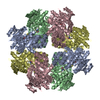
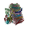
| Title | Crystal structure of P. aeruginosa geranyl-CoA carboxylase (GCC), beta subunit | ||||||||||||
 Components Components | Geranyl-CoA carboxylase, beta-subunit | ||||||||||||
 Keywords Keywords |  LIGASE / Carboxylase / organic acid metabolism LIGASE / Carboxylase / organic acid metabolism | ||||||||||||
| Function / homology |  Function and homology informationgeranoyl-CoA carboxylase activity / terpene catabolic process / methylcrotonoyl-CoA carboxylase complex / L-leucine catabolic process / fatty acid biosynthetic process Function and homology informationgeranoyl-CoA carboxylase activity / terpene catabolic process / methylcrotonoyl-CoA carboxylase complex / L-leucine catabolic process / fatty acid biosynthetic processSimilarity search - Function | ||||||||||||
| Biological species |   Pseudomonas aeruginosa (bacteria) Pseudomonas aeruginosa (bacteria) | ||||||||||||
| Method | X-RAY DIFFRACTION / SYNCHROTRON / MOLECULAR REPLACEMENT / molecular replacement / Resolution: 2.4 Å | ||||||||||||
 Authors Authors | Huang, C.S. / Jurado, A.R. / Tong, L. | ||||||||||||
| Funding support |  United States, 3items United States, 3items
| ||||||||||||
 Citation Citation | Journal: Nat Commun / Year: 2015 Title: Structure and substrate selectivity of the 750-kDa alpha6beta6 holoenzyme of geranyl-CoA carboxylase Authors: Jurado, A.R. / Huang, C.S. / Zhang, X. / Zhou, Z.H. / Tong, L. | ||||||||||||
| History |
|
- Structure visualization
| Structure viewer | Molecule: MolmilJmol/JSmol |
|---|
- Downloads & links
-Download
| PDBx/mmCIF format | 5ikl.cif.gz | 281.2 KB | Display | PDBx/mmCIF format |
|---|---|---|---|---|
| PDB format | pdb5ikl.ent.gz | 229.4 KB | Display | PDB format |
| PDBx/mmJSON format | 5ikl.json.gz | Tree view | PDBx/mmJSON format | |
| Others |  Other downloads Other downloads |
-Validation report
| Arichive directory | https://data.pdbj.org/pub/pdb/validation_reports/ik/5iklftp://data.pdbj.org/pub/pdb/validation_reports/ik/5ikl | HTTPS FTP |
|---|
-Related structure data
| Similar structure data |
|---|


-Links
 PDBj
PDBj- Assembly
| Deposited unit | 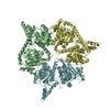
| ||||||||
|---|---|---|---|---|---|---|---|---|---|
| 1 | 
| ||||||||
| Unit cell |
|
-Components
| #1: Protein | Mass: 57263.141 Da / Num. of mol.: 3 Source method: isolated from a genetically manipulated source Source: (gene. exp.) Pseudomonas aeruginosa (strain ATCC 15692 / PAO1 / 1C / PRS 101 / LMG 12228) (bacteria)Strain: ATCC 15692 / PAO1 / 1C / PRS 101 / LMG 12228 / Gene: atuC, PA2888 / Production host: Escherichia coli (E. coli) / References: UniProt: Q9HZV9#2: Water | ChemComp-HOH / | Water Mass: 18.015 Da / Num. of mol.: 24 / Source method: isolated from a natural source / Formula: H2O Mass: 18.015 Da / Num. of mol.: 24 / Source method: isolated from a natural source / Formula: H2O |
|---|
-Experimental details
-Experiment
| Experiment | Method: X-RAY DIFFRACTION / Number of used crystals: 1 |
|---|
- Sample preparation
| Crystal | Density Matthews: 2.59 Å3/Da / Density % sol: 52.44 % |
|---|---|
| Crystal grow | Temperature: 293 K / Method: vapor diffusion, hanging drop / pH: 8 / Details: 0.1M Tris-HCl (pH 8.0), 60% (v/v) MPD |
-Data collection
| Diffraction | Mean temperature: 100 K | ||||||||||||||||||||||||||||||||||||||||||||||||||||||||||||||||||
|---|---|---|---|---|---|---|---|---|---|---|---|---|---|---|---|---|---|---|---|---|---|---|---|---|---|---|---|---|---|---|---|---|---|---|---|---|---|---|---|---|---|---|---|---|---|---|---|---|---|---|---|---|---|---|---|---|---|---|---|---|---|---|---|---|---|---|---|
| Diffraction source | Source: SYNCHROTRON / Site: NSLS / Beamline: X29A / Wavelength: 1.075 Å | ||||||||||||||||||||||||||||||||||||||||||||||||||||||||||||||||||
| Detector | Type: ADSC QUANTUM 315 / Detector: CCD / Date: Oct 27, 2012 | ||||||||||||||||||||||||||||||||||||||||||||||||||||||||||||||||||
| Radiation | Protocol: SINGLE WAVELENGTH / Monochromatic (M) / Laue (L): M / Scattering type: x-ray | ||||||||||||||||||||||||||||||||||||||||||||||||||||||||||||||||||
| Radiation wavelength | Wavelength: 1.075 Å / Relative weight: 1 | ||||||||||||||||||||||||||||||||||||||||||||||||||||||||||||||||||
| Reflection | Resolution: 2.4→50 Å / Num. obs: 71918 / % possible obs: 98.6 % / Redundancy: 4.5 % / Rmerge(I) obs: 0.084 / Χ2: 1.035 / Net I/av σ(I): 15.707 / Net I/σ(I): 11.7 / Num. measured all: 322453 | ||||||||||||||||||||||||||||||||||||||||||||||||||||||||||||||||||
| Reflection shell | Diffraction-ID: 1 / Rejects: 0
|
-Phasing
| Phasing | Method: molecular replacement |
|---|
- Processing
| Software |
| |||||||||||||||||||||||||||||||||||||||||||||||||||||||||||||||||||||||||||
|---|---|---|---|---|---|---|---|---|---|---|---|---|---|---|---|---|---|---|---|---|---|---|---|---|---|---|---|---|---|---|---|---|---|---|---|---|---|---|---|---|---|---|---|---|---|---|---|---|---|---|---|---|---|---|---|---|---|---|---|---|---|---|---|---|---|---|---|---|---|---|---|---|---|---|---|---|
| Refinement | Method to determine structure: MOLECULAR REPLACEMENT / Resolution: 2.4→47.195 Å / Cor.coef. Fo:Fc: 0.937 / Cor.coef. Fo:Fc free: 0.898 / SU B: 9.963 / SU ML: 0.228 / Cross valid method: THROUGHOUT / σ(F): 0 / ESU R: 0.364 / ESU R Free: 0.279 / Stereochemistry target values: MAXIMUM LIKELIHOOD Details: HYDROGENS HAVE BEEN ADDED IN THE RIDING POSITIONS U VALUES : REFINED INDIVIDUALLY
| |||||||||||||||||||||||||||||||||||||||||||||||||||||||||||||||||||||||||||
| Solvent computation | Ion probe radii: 0.8 Å / Shrinkage radii: 0.8 Å / VDW probe radii: 1.2 Å / Solvent model: MASK | |||||||||||||||||||||||||||||||||||||||||||||||||||||||||||||||||||||||||||
| Displacement parameters | Biso max: 201.29 Å2 / Biso mean: 65.984 Å2 / Biso min: 30.88 Å2
| |||||||||||||||||||||||||||||||||||||||||||||||||||||||||||||||||||||||||||
| Refinement step | Cycle: final / Resolution: 2.4→47.195 Å
| |||||||||||||||||||||||||||||||||||||||||||||||||||||||||||||||||||||||||||
| Refine LS restraints |
| |||||||||||||||||||||||||||||||||||||||||||||||||||||||||||||||||||||||||||
| LS refinement shell | Resolution: 2.401→2.463 Å / Total num. of bins used: 20
|
