 Movie
Movie Controller

[English] 日本語
 Yorodumi
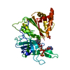
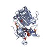
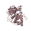
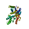
- PDB-5ih6: Human Casein Kinase 1 isoform delta (kinase domain) in complex wi... -
+ Open data

- Basic information
| Entry | Database: PDB / ID: 5ih6 | ||||||
|---|---|---|---|---|---|---|---|
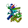
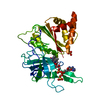
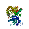
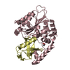
| Title | Human Casein Kinase 1 isoform delta (kinase domain) in complex with Epiblastin A derivative | ||||||
 Components Components | Casein kinase I isoform delta | ||||||
 Keywords Keywords |  TRANSFERASE / Kinase domain / stem cell reprogramming / kinase inhibitor complex TRANSFERASE / Kinase domain / stem cell reprogramming / kinase inhibitor complex | ||||||
| Function / homology |  Function and homology information Function and homology informationpositive regulation of non-canonical Wnt signaling pathway / protein localization to Golgi apparatus / COPII vesicle coating / midbrain dopaminergic neuron differentiation / microtubule nucleation / protein localization to cilium / tau-protein kinase / non-motile cilium assembly / protein localization to centrosome / COPII-mediated vesicle transport ...positive regulation of non-canonical Wnt signaling pathway / protein localization to Golgi apparatus / COPII vesicle coating / midbrain dopaminergic neuron differentiation / microtubule nucleation / protein localization to cilium / tau-protein kinase / non-motile cilium assembly / protein localization to centrosome / COPII-mediated vesicle transport / tau-protein kinase activity / Golgi organization / Major pathway of rRNA processing in the nucleolus and cytosol / spindle assembly / Loss of Nlp from mitotic centrosomes / Loss of proteins required for interphase microtubule organization from the centrosome / Recruitment of mitotic centrosome proteins and complexes / Recruitment of NuMA to mitotic centrosomes / Anchoring of the basal body to the plasma membrane / endoplasmic reticulum-Golgi intermediate compartment membrane / AURKA Activation by TPX2 / cellular response to nerve growth factor stimulus / ciliary basal body / circadian regulation of gene expression / spindle microtubule / regulation of circadian rhythm / Wnt signaling pathway / spindle / endocytosis / Regulation of PLK1 Activity at G2/M Transition / positive regulation of canonical Wnt signaling pathway / Circadian Clock / positive regulation of proteasomal ubiquitin-dependent protein catabolic process / non-specific serine/threonine protein kinase / protein kinase activity / cadherin binding / positive regulation of protein phosphorylation / protein phosphorylation / protein serine kinase activity / protein serine/threonine kinase activity / centrosome / perinuclear region of cytoplasm / Golgi apparatus / signal transduction / nucleoplasm / ATP binding / nucleus / plasma membrane / cytosolSimilarity search - Function | ||||||
| Biological species |  Homo sapiens (human) Homo sapiens (human) | ||||||
| Method | X-RAY DIFFRACTION / SYNCHROTRON / MOLECULAR REPLACEMENT / molecular replacement / Resolution: 2.3 Å | ||||||
 Authors Authors | Ursu, A. / Illich, D.J. / Takemoto, Y. / Porfetye, A.T. / Zhang, M. / Brockmeyer, A. / Janning, P. / Watanabe, N. / Osada, H. / Vetter, I.R. ...Ursu, A. / Illich, D.J. / Takemoto, Y. / Porfetye, A.T. / Zhang, M. / Brockmeyer, A. / Janning, P. / Watanabe, N. / Osada, H. / Vetter, I.R. / Ziegler, S. / Schoeler, H.R. / Waldmann, H. | ||||||
 Citation Citation | Journal: Cell Chem Biol / Year: 2016 Title: Epiblastin A Induces Reprogramming of Epiblast Stem Cells Into Embryonic Stem Cells by Inhibition of Casein Kinase 1. Authors: Ursu, A. / Illich, D.J. / Takemoto, Y. / Porfetye, A.T. / Zhang, M. / Brockmeyer, A. / Janning, P. / Watanabe, N. / Osada, H. / Vetter, I.R. / Ziegler, S. / Scholer, H.R. / Waldmann, H. | ||||||
| History |
|
- Structure visualization
| Structure viewer | Molecule: MolmilJmol/JSmol |
|---|
- Downloads & links
-Download
| PDBx/mmCIF format | 5ih6.cif.gz | 74.8 KB | Display | PDBx/mmCIF format |
|---|---|---|---|---|
| PDB format | pdb5ih6.ent.gz | 55.2 KB | Display | PDB format |
| PDBx/mmJSON format | 5ih6.json.gz | Tree view | PDBx/mmJSON format | |
| Others |  Other downloads Other downloads |
-Validation report
| Arichive directory | https://data.pdbj.org/pub/pdb/validation_reports/ih/5ih6ftp://data.pdbj.org/pub/pdb/validation_reports/ih/5ih6 | HTTPS FTP |
|---|
-Related structure data
| Related structure data |  5ih4SC 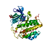 5ih5C S: Starting model for refinement C: citing same article ( |
|---|---|
| Similar structure data |


-Links
 PDBj
PDBj


- Assembly
| Deposited unit | 
| ||||||||
|---|---|---|---|---|---|---|---|---|---|
| 1 |
| ||||||||
| Unit cell |
|
-Components
-Protein , 1 types, 1 molecules A
| #1: Protein | Mass: 34206.531 Da / Num. of mol.: 1 Source method: isolated from a genetically manipulated source Details: N-terminal cloning artifact: GP / Source: (gene. exp.) Homo sapiens (human) / Gene: CSNK1D, HCKID / Production host:  Escherichia coli BL21(DE3) (bacteria) / Variant (production host): RIL Escherichia coli BL21(DE3) (bacteria) / Variant (production host): RILReferences: UniProt: P48730, non-specific serine/threonine protein kinase, tau-protein kinase |
|---|
-Non-polymers , 5 types, 27 molecules 


| #2: Chemical | ChemComp-ZN /  Mass: 65.409 Da / Num. of mol.: 1 / Source method: obtained synthetically / Formula: Zn Mass: 65.409 Da / Num. of mol.: 1 / Source method: obtained synthetically / Formula: Zn | ||||
|---|---|---|---|---|---|
| #3: Chemical | ChemComp-SRT /  Mass: 150.087 Da / Num. of mol.: 1 / Source method: obtained synthetically / Formula: C4H6O6 Mass: 150.087 Da / Num. of mol.: 1 / Source method: obtained synthetically / Formula: C4H6O6 | ||||
| #4: Chemical | Sulfate Mass: 96.063 Da / Num. of mol.: 2 / Source method: obtained synthetically / Formula: SO4 Mass: 96.063 Da / Num. of mol.: 2 / Source method: obtained synthetically / Formula: SO4#5: Chemical | ChemComp-AUG / |  Mass: 332.159 Da / Num. of mol.: 1 / Source method: obtained synthetically / Formula: C12H10BrN7 Mass: 332.159 Da / Num. of mol.: 1 / Source method: obtained synthetically / Formula: C12H10BrN7#6: Water | ChemComp-HOH / | WaterMass: 18.015 Da / Num. of mol.: 22 / Source method: isolated from a natural source / Formula: H2O |
-Experimental details
-Experiment
| Experiment | Method: X-RAY DIFFRACTION / Number of used crystals: 1 |
|---|
- Sample preparation
| Crystal | Density Matthews: 2.79 Å3/Da / Density % sol: 55.99 % |
|---|---|
| Crystal grow | Temperature: 277 K / Method: vapor diffusion, sitting drop Details: 0.1 M Li2SO4, 0.7 - 0.8 M Na-K tartrate, 0.1 M CHES PH range: 9.3-9.5 |
-Data collection
| Diffraction | Mean temperature: 100 K | |||||||||||||||||||||||||||||||||||||||||||||||||||||||||||||||||||||||||||||||||||||||||||||||||||||||||||||||||||||||||||||||||||||||||||||||||||||||||||||||||||||||||||||||||||||||||||||||||||||||||||||||||||||||||||||||||||||||
|---|---|---|---|---|---|---|---|---|---|---|---|---|---|---|---|---|---|---|---|---|---|---|---|---|---|---|---|---|---|---|---|---|---|---|---|---|---|---|---|---|---|---|---|---|---|---|---|---|---|---|---|---|---|---|---|---|---|---|---|---|---|---|---|---|---|---|---|---|---|---|---|---|---|---|---|---|---|---|---|---|---|---|---|---|---|---|---|---|---|---|---|---|---|---|---|---|---|---|---|---|---|---|---|---|---|---|---|---|---|---|---|---|---|---|---|---|---|---|---|---|---|---|---|---|---|---|---|---|---|---|---|---|---|---|---|---|---|---|---|---|---|---|---|---|---|---|---|---|---|---|---|---|---|---|---|---|---|---|---|---|---|---|---|---|---|---|---|---|---|---|---|---|---|---|---|---|---|---|---|---|---|---|---|---|---|---|---|---|---|---|---|---|---|---|---|---|---|---|---|---|---|---|---|---|---|---|---|---|---|---|---|---|---|---|---|---|---|---|---|---|---|---|---|---|---|---|---|---|---|---|---|---|
| Diffraction source | Source: SYNCHROTRON / Site: SLS  / Beamline: X10SA / Wavelength: 1.76995 Å / Beamline: X10SA / Wavelength: 1.76995 Å | |||||||||||||||||||||||||||||||||||||||||||||||||||||||||||||||||||||||||||||||||||||||||||||||||||||||||||||||||||||||||||||||||||||||||||||||||||||||||||||||||||||||||||||||||||||||||||||||||||||||||||||||||||||||||||||||||||||||
| Detector | Type: DECTRIS PILATUS 6M / Detector: PIXEL / Date: Mar 19, 2015 | |||||||||||||||||||||||||||||||||||||||||||||||||||||||||||||||||||||||||||||||||||||||||||||||||||||||||||||||||||||||||||||||||||||||||||||||||||||||||||||||||||||||||||||||||||||||||||||||||||||||||||||||||||||||||||||||||||||||
| Radiation | Protocol: SINGLE WAVELENGTH / Monochromatic (M) / Laue (L): M / Scattering type: x-ray | |||||||||||||||||||||||||||||||||||||||||||||||||||||||||||||||||||||||||||||||||||||||||||||||||||||||||||||||||||||||||||||||||||||||||||||||||||||||||||||||||||||||||||||||||||||||||||||||||||||||||||||||||||||||||||||||||||||||
| Radiation wavelength | Wavelength: 1.76995 Å / Relative weight: 1 | |||||||||||||||||||||||||||||||||||||||||||||||||||||||||||||||||||||||||||||||||||||||||||||||||||||||||||||||||||||||||||||||||||||||||||||||||||||||||||||||||||||||||||||||||||||||||||||||||||||||||||||||||||||||||||||||||||||||
| Reflection | Resolution: 2.3→37.8 Å / Num. obs: 17269 / % possible obs: 98.3 % / Observed criterion σ(I): -3 / Redundancy: 14.1 % / Biso Wilson estimate: 62.054 Å2 / CC1/2: 0.999 / Rmerge(I) obs: 0.115 / Rrim(I) all: 0.119 / Χ2: 1.126 / Net I/σ(I): 12.49 / Num. measured all: 243801 | |||||||||||||||||||||||||||||||||||||||||||||||||||||||||||||||||||||||||||||||||||||||||||||||||||||||||||||||||||||||||||||||||||||||||||||||||||||||||||||||||||||||||||||||||||||||||||||||||||||||||||||||||||||||||||||||||||||||
| Reflection shell | Diffraction-ID: 1 / Rejects: 0
|
-Phasing
| Phasing | Method: molecular replacement |
|---|
- Processing
| Software |
| |||||||||||||||||||||||||||||||||||||||||||||||||||||||||||||||||||||||||||
|---|---|---|---|---|---|---|---|---|---|---|---|---|---|---|---|---|---|---|---|---|---|---|---|---|---|---|---|---|---|---|---|---|---|---|---|---|---|---|---|---|---|---|---|---|---|---|---|---|---|---|---|---|---|---|---|---|---|---|---|---|---|---|---|---|---|---|---|---|---|---|---|---|---|---|---|---|
| Refinement | Method to determine structure: MOLECULAR REPLACEMENT Starting model: 5IH4 Resolution: 2.3→37.8 Å / Cor.coef. Fo:Fc: 0.952 / Cor.coef. Fo:Fc free: 0.952 / SU B: 15.112 / SU ML: 0.307 / Cross valid method: THROUGHOUT / σ(F): 0 / ESU R: 0.327 / ESU R Free: 0.234 / Stereochemistry target values: MAXIMUM LIKELIHOOD Details: HYDROGENS HAVE BEEN ADDED IN THE RIDING POSITIONS U VALUES : REFINED INDIVIDUALLY
| |||||||||||||||||||||||||||||||||||||||||||||||||||||||||||||||||||||||||||
| Solvent computation | Ion probe radii: 0.8 Å / Shrinkage radii: 0.8 Å / VDW probe radii: 1.2 Å / Solvent model: MASK | |||||||||||||||||||||||||||||||||||||||||||||||||||||||||||||||||||||||||||
| Displacement parameters | Biso max: 218.68 Å2 / Biso mean: 89.694 Å2 / Biso min: 35.13 Å2
| |||||||||||||||||||||||||||||||||||||||||||||||||||||||||||||||||||||||||||
| Refinement step | Cycle: final / Resolution: 2.3→37.8 Å
| |||||||||||||||||||||||||||||||||||||||||||||||||||||||||||||||||||||||||||
| Refine LS restraints |
| |||||||||||||||||||||||||||||||||||||||||||||||||||||||||||||||||||||||||||
| LS refinement shell | Resolution: 2.3→2.359 Å / Total num. of bins used: 20
|
