 Movie
Movie Controller

[English] 日本語
 Yorodumi
- PDB-5iat: Mechanistic and Structural Analysis of Substrate Recognition and ... -
+ Open data

- Basic information
| Entry | Database: PDB / ID: 5iat | ||||||
|---|---|---|---|---|---|---|---|
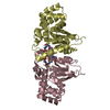
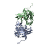
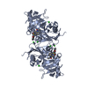
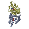
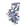
| Title | Mechanistic and Structural Analysis of Substrate Recognition and Cofactor Binding by an Unusual Bacterial Prolyl Hydroxylase - apo-BaP4H | ||||||
 Components Components | Procollagen-Proline Dioxygenase | ||||||
 Keywords Keywords | OXIDOREDUCTASE / P4H / Dioxygenase / Cupin / Fe(II)/alpha-ketoglutarate | ||||||
| Function / homology |  Function and homology information Function and homology informationprocollagen-proline 4-dioxygenase activity / L-ascorbic acid binding / dioxygenase activity / oxidoreductase activity, acting on paired donors, with incorporation or reduction of molecular oxygen / iron ion bindingSimilarity search - Function | ||||||
| Biological species |  Bacillus anthracis (anthrax bacterium) Bacillus anthracis (anthrax bacterium) | ||||||
| Method | X-RAY DIFFRACTION / SYNCHROTRON / MOLECULAR REPLACEMENT / molecular replacement / Resolution: 1.67 Å | ||||||
 Authors Authors | Schnicker, N.J. / Dey, M. | ||||||
| Funding support |  United States, 1items United States, 1items
| ||||||
 Citation Citation | Journal: J.Biol.Chem. / Year: 2016 Title: Bacillus anthracis Prolyl 4-Hydroxylase Modifies Collagen-like Substrates in Asymmetric Patterns. Authors: Schnicker, N.J. / Dey, M. | ||||||
| History |
|
- Structure visualization
| Structure viewer | Molecule: MolmilJmol/JSmol |
|---|
- Downloads & links
-Download
| PDBx/mmCIF format | 5iat.cif.gz | 103.8 KB | Display | PDBx/mmCIF format |
|---|---|---|---|---|
| PDB format | pdb5iat.ent.gz | 76.3 KB | Display | PDB format |
| PDBx/mmJSON format | 5iat.json.gz | Tree view | PDBx/mmJSON format | |
| Others |  Other downloads Other downloads |
-Validation report
| Arichive directory | https://data.pdbj.org/pub/pdb/validation_reports/ia/5iatftp://data.pdbj.org/pub/pdb/validation_reports/ia/5iat | HTTPS FTP |
|---|
-Related structure data
| Related structure data |  5iavC 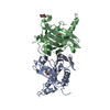 5iaxC  3itqS S: Starting model for refinement C: citing same article ( |
|---|---|
| Similar structure data |


-Links
 PDBj
PDBj
- Assembly
| Deposited unit | 
| ||||||||
|---|---|---|---|---|---|---|---|---|---|
| 1 |
| ||||||||
| Unit cell |
|
-Components
| #1: Protein | Mass: 24708.592 Da / Num. of mol.: 2 / Fragment: UNP residues 18-232 Source method: isolated from a genetically manipulated source Source: (gene. exp.) Bacillus anthracis (anthrax bacterium) / Gene: BASH2_01493 / Production host: Escherichia coli (E. coli) / References: UniProt: A0A0F7R8C5, UniProt: A0A4Y1WAP5*PLUS#2: Chemical | Glycerol  Mass: 92.094 Da / Num. of mol.: 2 / Source method: obtained synthetically / Formula: C3H8O3 Mass: 92.094 Da / Num. of mol.: 2 / Source method: obtained synthetically / Formula: C3H8O3#3: Chemical | Diethylene glycol  Mass: 106.120 Da / Num. of mol.: 2 / Source method: obtained synthetically / Formula: C4H10O3 Mass: 106.120 Da / Num. of mol.: 2 / Source method: obtained synthetically / Formula: C4H10O3#4: Water | ChemComp-HOH / | Water Mass: 18.015 Da / Num. of mol.: 396 / Source method: isolated from a natural source / Formula: H2O Mass: 18.015 Da / Num. of mol.: 396 / Source method: isolated from a natural source / Formula: H2O |
|---|
-Experimental details
-Experiment
| Experiment | Method: X-RAY DIFFRACTION / Number of used crystals: 1 |
|---|
- Sample preparation
| Crystal | Density Matthews: 2.17 Å3/Da / Density % sol: 43.31 % |
|---|---|
| Crystal grow | Temperature: 293 K / Method: vapor diffusion, hanging drop / pH: 6 / Details: 40 mM KH2PO4 pH 6, 14 % PEG 8000, 20 % Glycerol |
-Data collection
| Diffraction | Mean temperature: 100 K |
|---|---|
| Diffraction source | Source: SYNCHROTRON / Site: ALS / Beamline: 4.2.2 / Wavelength: 1 Å |
| Detector | Type: NOIR-1 / Detector: CCD / Date: Jun 21, 2013 |
| Radiation | Protocol: SINGLE WAVELENGTH / Monochromatic (M) / Laue (L): M / Scattering type: x-ray |
| Radiation wavelength | Wavelength: 1 Å / Relative weight: 1 |
| Reflection | Resolution: 1.67→33.28 Å / Num. obs: 50418 / % possible obs: 99.5 % / Redundancy: 6.9 % / Rmerge(I) obs: 0.042 / Net I/σ(I): 27.1 |
| Reflection shell | Resolution: 1.67→1.76 Å / Redundancy: 4.6 % / Rmerge(I) obs: 0.46 / Mean I/σ(I) obs: 3.2 / Num. measured obs: 6990 / % possible all: 96.5 |
-Phasing
| Phasing | Method: molecular replacement |
|---|
- Processing
| Software |
| |||||||||||||||||||||||||||||||||||||||||||||||||||||||||||||||||||||||||||||||||||||||||||||||||||||||||||||||||||||||||||||||||||||
|---|---|---|---|---|---|---|---|---|---|---|---|---|---|---|---|---|---|---|---|---|---|---|---|---|---|---|---|---|---|---|---|---|---|---|---|---|---|---|---|---|---|---|---|---|---|---|---|---|---|---|---|---|---|---|---|---|---|---|---|---|---|---|---|---|---|---|---|---|---|---|---|---|---|---|---|---|---|---|---|---|---|---|---|---|---|---|---|---|---|---|---|---|---|---|---|---|---|---|---|---|---|---|---|---|---|---|---|---|---|---|---|---|---|---|---|---|---|---|---|---|---|---|---|---|---|---|---|---|---|---|---|---|---|---|
| Refinement | Method to determine structure: MOLECULAR REPLACEMENT Starting model: 3ITQ Resolution: 1.67→33.279 Å / SU ML: 0.16 / Cross valid method: FREE R-VALUE / σ(F): 1.35 / Phase error: 20.86
| |||||||||||||||||||||||||||||||||||||||||||||||||||||||||||||||||||||||||||||||||||||||||||||||||||||||||||||||||||||||||||||||||||||
| Solvent computation | Shrinkage radii: 0.9 Å / VDW probe radii: 1.11 Å | |||||||||||||||||||||||||||||||||||||||||||||||||||||||||||||||||||||||||||||||||||||||||||||||||||||||||||||||||||||||||||||||||||||
| Displacement parameters | Biso max: 100.33 Å2 / Biso mean: 23.4144 Å2 / Biso min: 6.39 Å2 | |||||||||||||||||||||||||||||||||||||||||||||||||||||||||||||||||||||||||||||||||||||||||||||||||||||||||||||||||||||||||||||||||||||
| Refinement step | Cycle: final / Resolution: 1.67→33.279 Å
| |||||||||||||||||||||||||||||||||||||||||||||||||||||||||||||||||||||||||||||||||||||||||||||||||||||||||||||||||||||||||||||||||||||
| Refine LS restraints |
| |||||||||||||||||||||||||||||||||||||||||||||||||||||||||||||||||||||||||||||||||||||||||||||||||||||||||||||||||||||||||||||||||||||
| LS refinement shell | Refine-ID: X-RAY DIFFRACTION / Total num. of bins used: 18
|
