 Movie
Movie Controller

[English] 日本語
 Yorodumi
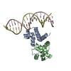
- PDB-5i7m: Crystal structure of Y345F mutant of human primase p58 iron-sulfu... -
+ Open data

- Basic information
| Entry | Database: PDB / ID: 5i7m | ||||||||||||||||||||||||
|---|---|---|---|---|---|---|---|---|---|---|---|---|---|---|---|---|---|---|---|---|---|---|---|---|---|
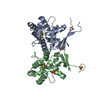
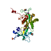
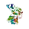
| Title | Crystal structure of Y345F mutant of human primase p58 iron-sulfur cluster domain | ||||||||||||||||||||||||
 Components Components | DNA primase large subunit Primase Primase | ||||||||||||||||||||||||
 Keywords Keywords | REPLICATION / DNA replication / primase / [4Fe-4S] / p58C | ||||||||||||||||||||||||
| Function / homology |  Function and homology information Function and homology informationpositive regulation of DNA primase activity / DNA replication initiation / DNA/RNA hybrid binding / Telomere C-strand synthesis initiation / Inhibition of replication initiation of damaged DNA by RB1/E2F1 / alpha DNA polymerase:primase complex / Polymerase switching / Processive synthesis on the lagging strand / Removal of the Flap Intermediate / Polymerase switching on the C-strand of the telomere ...positive regulation of DNA primase activity / DNA replication initiation / DNA/RNA hybrid binding / Telomere C-strand synthesis initiation / Inhibition of replication initiation of damaged DNA by RB1/E2F1 / alpha DNA polymerase:primase complex / Polymerase switching / Processive synthesis on the lagging strand / Removal of the Flap Intermediate / Polymerase switching on the C-strand of the telomere / DNA replication, synthesis of primer / Activation of the pre-replicative complex / DNA replication initiation / Defective pyroptosis / 4 iron, 4 sulfur cluster binding / DNA binding / nucleoplasm / metal ion bindingSimilarity search - Function | ||||||||||||||||||||||||
| Biological species |  Homo sapiens (human) Homo sapiens (human) | ||||||||||||||||||||||||
| Method | X-RAY DIFFRACTION / SYNCHROTRON / MOLECULAR REPLACEMENT / molecular replacement / Resolution: 1.93 Å | ||||||||||||||||||||||||
 Authors Authors | Salay, L.E. / Thompson, M.K. / Chazin, W.J. | ||||||||||||||||||||||||
| Funding support |  United States, 7items United States, 7items
| ||||||||||||||||||||||||
 Citation Citation | Journal: Science / Year: 2017 Title: The [4Fe4S] cluster of human DNA primase functions as a redox switch using DNA charge transport. Authors: O'Brien, E. / Holt, M.E. / Thompson, M.K. / Salay, L.E. / Ehlinger, A.C. / Chazin, W.J. / Barton, J.K. | ||||||||||||||||||||||||
| History |
|
- Structure visualization
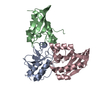
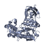
| Structure viewer | Molecule: MolmilJmol/JSmol |
|---|
- Downloads & links
-Download
| PDBx/mmCIF format | 5i7m.cif.gz | 84.2 KB | Display | PDBx/mmCIF format |
|---|---|---|---|---|
| PDB format | pdb5i7m.ent.gz | 61 KB | Display | PDB format |
| PDBx/mmJSON format | 5i7m.json.gz | Tree view | PDBx/mmJSON format | |
| Others |  Other downloads Other downloads |
-Validation report
| Arichive directory | https://data.pdbj.org/pub/pdb/validation_reports/i7/5i7mftp://data.pdbj.org/pub/pdb/validation_reports/i7/5i7m | HTTPS FTP |
|---|
-Related structure data
| Related structure data | 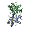 5dqoC 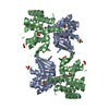 3l9qS S: Starting model for refinement C: citing same article ( |
|---|---|
| Similar structure data |


-Links
 PDBj
PDBj


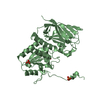
- Assembly
| Deposited unit | 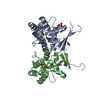
| ||||||||
|---|---|---|---|---|---|---|---|---|---|
| 1 |
| ||||||||
| Unit cell |
|
-Components
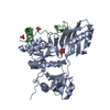
| #1: Protein | Primase / Primase regulatory subunit / DNA primase 58 kDa subunit / p58 Mass: 21709.867 Da / Num. of mol.: 2 / Fragment: iron-sulfur cluster domain (UNP residues 272-457) / Mutation: Y345F Source method: isolated from a genetically manipulated source Source: (gene. exp.) Homo sapiens (human) / Gene: PRIM2, PRIM2A / Production host:  Escherichia coli BL21(DE3) (bacteria) Escherichia coli BL21(DE3) (bacteria)References: UniProt: P49643, Transferases; Transferring phosphorus-containing groups; Nucleotidyltransferases#2: Chemical | Iron–sulfur cluster  Mass: 351.640 Da / Num. of mol.: 2 / Source method: obtained synthetically / Formula: Fe4S4 Mass: 351.640 Da / Num. of mol.: 2 / Source method: obtained synthetically / Formula: Fe4S4#3: Chemical | ChemComp-SO4 / | Sulfate  Mass: 96.063 Da / Num. of mol.: 1 / Source method: obtained synthetically / Formula: SO4 Mass: 96.063 Da / Num. of mol.: 1 / Source method: obtained synthetically / Formula: SO4#4: Water | ChemComp-HOH / | Water Mass: 18.015 Da / Num. of mol.: 103 / Source method: isolated from a natural source / Formula: H2O Mass: 18.015 Da / Num. of mol.: 103 / Source method: isolated from a natural source / Formula: H2O |
|---|
-Experimental details
-Experiment
| Experiment | Method: X-RAY DIFFRACTION / Number of used crystals: 1 |
|---|
- Sample preparation
| Crystal | Density Matthews: 2.75 Å3/Da / Density % sol: 55.3 % |
|---|---|
| Crystal grow | Temperature: 277 K / Method: vapor diffusion, hanging drop / pH: 6.5 Details: 75 mg/mL p58C Y345F in 20 mM MES, pH 6.5, 50 mM sodium chloride, reservoir solution: 100 mM Tris, pH 8.5, 150 mM lithium sulfate, 18% PEG3350 |
-Data collection
| Diffraction | Mean temperature: 100 K | |||||||||||||||||||||||||||||||||||||||||||||||||||||||||||||||||||||||||||||||||||||||||||||||||||||||||||||||||||||||||||||||||||||||||||||||||||||||||||||||||||||||||||||||||||||||||||||
|---|---|---|---|---|---|---|---|---|---|---|---|---|---|---|---|---|---|---|---|---|---|---|---|---|---|---|---|---|---|---|---|---|---|---|---|---|---|---|---|---|---|---|---|---|---|---|---|---|---|---|---|---|---|---|---|---|---|---|---|---|---|---|---|---|---|---|---|---|---|---|---|---|---|---|---|---|---|---|---|---|---|---|---|---|---|---|---|---|---|---|---|---|---|---|---|---|---|---|---|---|---|---|---|---|---|---|---|---|---|---|---|---|---|---|---|---|---|---|---|---|---|---|---|---|---|---|---|---|---|---|---|---|---|---|---|---|---|---|---|---|---|---|---|---|---|---|---|---|---|---|---|---|---|---|---|---|---|---|---|---|---|---|---|---|---|---|---|---|---|---|---|---|---|---|---|---|---|---|---|---|---|---|---|---|---|---|---|---|---|---|
| Diffraction source | Source: SYNCHROTRON / Site: APS / Beamline: 21-ID-G / Wavelength: 0.987 Å | |||||||||||||||||||||||||||||||||||||||||||||||||||||||||||||||||||||||||||||||||||||||||||||||||||||||||||||||||||||||||||||||||||||||||||||||||||||||||||||||||||||||||||||||||||||||||||||
| Detector | Type: MARMOSAIC 300 mm CCD / Detector: CCD / Date: Feb 6, 2016 | |||||||||||||||||||||||||||||||||||||||||||||||||||||||||||||||||||||||||||||||||||||||||||||||||||||||||||||||||||||||||||||||||||||||||||||||||||||||||||||||||||||||||||||||||||||||||||||
| Radiation | Monochromator: diamond(111) / Protocol: SINGLE WAVELENGTH / Monochromatic (M) / Laue (L): M / Scattering type: x-ray | |||||||||||||||||||||||||||||||||||||||||||||||||||||||||||||||||||||||||||||||||||||||||||||||||||||||||||||||||||||||||||||||||||||||||||||||||||||||||||||||||||||||||||||||||||||||||||||
| Radiation wavelength | Wavelength: 0.987 Å / Relative weight: 1 | |||||||||||||||||||||||||||||||||||||||||||||||||||||||||||||||||||||||||||||||||||||||||||||||||||||||||||||||||||||||||||||||||||||||||||||||||||||||||||||||||||||||||||||||||||||||||||||
| Reflection | Resolution: 1.93→80.66 Å / Num. obs: 34870 / % possible obs: 99.9 % / Redundancy: 4.2 % / Rmerge(I) obs: 0.087 / Rpim(I) all: 0.048 / Rrim(I) all: 0.099 / Χ2: 1.475 / Net I/av σ(I): 18.692 / Net I/σ(I): 6.9 / Num. measured all: 144740 | |||||||||||||||||||||||||||||||||||||||||||||||||||||||||||||||||||||||||||||||||||||||||||||||||||||||||||||||||||||||||||||||||||||||||||||||||||||||||||||||||||||||||||||||||||||||||||||
| Reflection shell | Diffraction-ID: 1 / Rejects: 0
|
-Phasing
| Phasing | Method: molecular replacement | |||||||||
|---|---|---|---|---|---|---|---|---|---|---|
| Phasing MR | Model details: Phaser MODE: MR_AUTO
|
- Processing
| Software |
| ||||||||||||||||||||||||
|---|---|---|---|---|---|---|---|---|---|---|---|---|---|---|---|---|---|---|---|---|---|---|---|---|---|
| Refinement | Method to determine structure: MOLECULAR REPLACEMENT Starting model: PDB entry 3L9Q Resolution: 1.93→80.66 Å / Cor.coef. Fo:Fc: 0.95 / Cor.coef. Fo:Fc free: 0.934 / SU B: 0.003 / SU ML: 0 / Cross valid method: THROUGHOUT / σ(F): 0 / ESU R: 0.12 / ESU R Free: 0.142 / Stereochemistry target values: MAXIMUM LIKELIHOOD / Details: HYDROGENS HAVE BEEN ADDED IN THE RIDING POSITIONS
| ||||||||||||||||||||||||
| Solvent computation | Ion probe radii: 0.8 Å / Shrinkage radii: 0.8 Å / VDW probe radii: 1.2 Å / Solvent model: MASK | ||||||||||||||||||||||||
| Displacement parameters | Biso max: 69.72 Å2 / Biso mean: 29.392 Å2 / Biso min: 14.98 Å2
| ||||||||||||||||||||||||
| Refinement step | Cycle: final / Resolution: 1.93→80.66 Å
| ||||||||||||||||||||||||
| LS refinement shell | Resolution: 1.93→1.98 Å / Total num. of bins used: 20
|
