Movie
Movie Controller


+ Open data

- Basic information
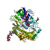
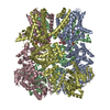
| Entry | Database: PDB / ID: 5i68 | ||||||
|---|---|---|---|---|---|---|---|
| Title | Alcohol oxidase from Pichia pastoris | ||||||
 Components Components | Alcohol oxidase 1 | ||||||
 Keywords Keywords | OXIDOREDUCTASE / alcohol oxidase peroxisome | ||||||
| Function / homology |  Function and homology information Function and homology informationmethane catabolic process / alcohol oxidase activity / alcohol oxidase / methanol metabolic process / peroxisomal matrix / flavin adenine dinucleotide bindingSimilarity search - Function | ||||||
| Biological species |  Komagataella pastoris (fungus) Komagataella pastoris (fungus) | ||||||
| Method | ELECTRON MICROSCOPY / single particle reconstruction / cryo EM / Resolution: 3.37 Å | ||||||
 Authors Authors | Vonck, J. / Mills, D.J. / Parcej, D.N. | ||||||
 Citation Citation | Journal: PLoS One / Year: 2016 Title: Structure of Alcohol Oxidase from Pichia pastoris by Cryo-Electron Microscopy. Authors: Janet Vonck / David N Parcej / Deryck J Mills /  Abstract: The first step in methanol metabolism in methylotrophic yeasts, the oxidation of methanol and higher alcohols with molecular oxygen to formaldehyde and hydrogen peroxide, is catalysed by alcohol ...The first step in methanol metabolism in methylotrophic yeasts, the oxidation of methanol and higher alcohols with molecular oxygen to formaldehyde and hydrogen peroxide, is catalysed by alcohol oxidase (AOX), a 600-kDa homo-octamer containing eight FAD cofactors. When these yeasts are grown with methanol as the carbon source, AOX forms large crystalline arrays in peroxisomes. We determined the structure of AOX by cryo-electron microscopy at a resolution of 3.4 Å. All residues of the 662-amino acid polypeptide as well as the FAD are well resolved. AOX shows high structural homology to other members of the GMC family of oxidoreductases, which share a conserved FAD binding domain, but have different substrate specificities. The preference of AOX for small alcohols is explained by the presence of conserved bulky aromatic residues near the active site. Compared to the other GMC enzymes, AOX contains a large number of amino acid inserts, the longest being 75 residues. These segments are found at the periphery of the monomer and make extensive inter-subunit contacts which are responsible for the very stable octamer. A short surface helix forms contacts between two octamers, explaining the tendency of AOX to form crystals in the peroxisomes. | ||||||
| History |
|
- Structure visualization
| Movie |
Movie viewer |
|---|---|
| Structure viewer | Molecule: MolmilJmol/JSmol |
- Downloads & links
-Download
| PDBx/mmCIF format | 5i68.cif.gz | 129.6 KB | Display | PDBx/mmCIF format |
|---|---|---|---|---|
| PDB format | pdb5i68.ent.gz | 103.2 KB | Display | PDB format |
| PDBx/mmJSON format | 5i68.json.gz | Tree view | PDBx/mmJSON format | |
| Others |  Other downloads Other downloads |
-Validation report
| Arichive directory | https://data.pdbj.org/pub/pdb/validation_reports/i6/5i68ftp://data.pdbj.org/pub/pdb/validation_reports/i6/5i68 | HTTPS FTP |
|---|
-Related structure data
| Related structure data |  8072MC M: map data used to model this data C: citing same article ( |
|---|---|
| Similar structure data |

-Links
 PDBj
PDBj
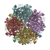
- Assembly
| Deposited unit | 
|
|---|---|
| 1 | x 8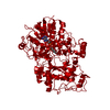
|
| Symmetry | Point symmetry: (Schoenflies symbol: D4 (2x4 fold dihedral)) |
-Components
| #1: Protein | / AOX 1 / Methanol oxidase 1 / MOX 1 Mass: 73992.195 Da / Num. of mol.: 1 / Source method: isolated from a natural source / Source: (natural) Komagataella pastoris (fungus)References: UniProt: F2QY27, UniProt: P04842*PLUS, alcohol oxidase |
|---|---|
| #2: Chemical | ChemComp-MG /   Mass: 24.305 Da / Num. of mol.: 1 / Source method: obtained synthetically / Formula: Mg Mass: 24.305 Da / Num. of mol.: 1 / Source method: obtained synthetically / Formula: Mg |
| #3: Chemical | ChemComp-FAD / Flavin adenine dinucleotide  Mass: 785.550 Da / Num. of mol.: 1 / Source method: obtained synthetically / Formula: C27H33N9O15P2 / Comment: FAD*YM Mass: 785.550 Da / Num. of mol.: 1 / Source method: obtained synthetically / Formula: C27H33N9O15P2 / Comment: FAD*YM |
-Experimental details
-Experiment
| Experiment | Method: ELECTRON MICROSCOPY |
|---|---|
| EM experiment | Aggregation state: PARTICLE / 3D reconstruction method: single particle reconstruction |
- Sample preparation
| Component | Name: alcohol oxidase / Type: COMPLEX / Entity ID: #1 / Source: NATURAL |
|---|---|
| Molecular weight | Value: 0.6 MDa / Experimental value: NO |
| Source (natural) | Organism: Komagataella pastoris (fungus) |
| Buffer solution | pH: 7.5 / Details: Potassium phosphate buffer, 50 mM |
| Specimen | Conc.: 0.7 mg/ml / Embedding applied: NO / Shadowing applied: NO / Staining applied: NO / Vitrification applied: YES / Details: This sample was monodisperse |
| Specimen support | Details: The grids had been cleaned in chloroform for 2 hrs. Grid material: COPPER / Grid mesh size: 400 divisions/in. / Grid type: Quantifoil R2/2 |
| Vitrification | Instrument: FEI VITROBOT MARK I / Cryogen name: ETHANE / Humidity: 70 % / Chamber temperature: 283 K / Details: blot for 11 seconds before plunging |
- Electron microscopy imaging
| Microscopy | Model: JEOL 3200FSC / Details: Data was collected manually |
|---|---|
| Electron gun | Electron source: FIELD EMISSION GUN / Accelerating voltage: 300 kV / Illumination mode: FLOOD BEAM |
| Electron lens | Mode: BRIGHT FIELDBright-field microscopy / Nominal magnification: 30000 X / Calibrated magnification: 43860 X / Calibrated defocus min: 600 nm / Calibrated defocus max: 2500 nm / Cs: 4.2 mm / Alignment procedure: COMA FREE |
| Specimen holder | Cryogen: NITROGEN / Specimen holder model: JEOL 3200FSC CRYOHOLDER |
| Image recording | Average exposure time: 6 sec. / Electron dose: 51 e/Å2 / Detector mode: COUNTING / Film or detector model: GATAN K2 SUMMIT (4k x 4k) |
| EM imaging optics | Energyfilter name: In-column Omega Filter / Energyfilter upper: 20 eV / Energyfilter lower: 0 eV |
| Image scans | Sampling size: 5 µm / Movie frames/image: 30 / Used frames/image: 2-21 |
- Processing
| EM software |
| ||||||||||||||||||||||||||||||||||||||||||||||||||
|---|---|---|---|---|---|---|---|---|---|---|---|---|---|---|---|---|---|---|---|---|---|---|---|---|---|---|---|---|---|---|---|---|---|---|---|---|---|---|---|---|---|---|---|---|---|---|---|---|---|---|---|
| Image processing | Details: The movie frames were aligned prior to particle picking and the images were binned 3x. | ||||||||||||||||||||||||||||||||||||||||||||||||||
| EM 3D crystal entity | ∠α: 90 ° / ∠β: 90 ° / ∠γ: 90 ° / A: 1 Å / B: 1 Å / C: 1 Å / Space group name: 1 / Space group num: 1 | ||||||||||||||||||||||||||||||||||||||||||||||||||
| CTF correction | Details: CTF was determined by CTFFIND3 inside the RELION software Type: PHASE FLIPPING ONLY | ||||||||||||||||||||||||||||||||||||||||||||||||||
| Particle selection | Num. of particles selected: 56544 | ||||||||||||||||||||||||||||||||||||||||||||||||||
| Symmetry | Point symmetry: D4 (2x4 fold dihedral) | ||||||||||||||||||||||||||||||||||||||||||||||||||
| 3D reconstruction | Resolution: 3.37 Å / Resolution method: FSC 0.143 CUT-OFF / Num. of particles: 49559 / Details: RELION was used for the reconstruction / Num. of class averages: 1 / Symmetry type: POINT | ||||||||||||||||||||||||||||||||||||||||||||||||||
| Atomic model building | B value: 147 / Protocol: AB INITIO MODEL / Space: REAL | ||||||||||||||||||||||||||||||||||||||||||||||||||
| Atomic model building |
|