 Movie
Movie Controller

[English] 日本語
 Yorodumi
- PDB-5i10: Crystal structure of spinosyn rhamnosyl 4'-O-methyltransferase sp... -
+ Open data

- Basic information
| Entry | Database: PDB / ID: 5i10 | ||||||
|---|---|---|---|---|---|---|---|
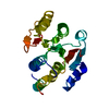
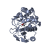
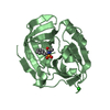
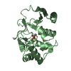
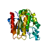
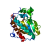
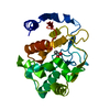
| Title | Crystal structure of spinosyn rhamnosyl 4'-O-methyltransferase spnh mutant T242Q from Saccharopolyspora Spinosa | ||||||
 Components Components | Probable O-methyltransferase | ||||||
 Keywords Keywords |  TRANSFERASE TRANSFERASE | ||||||
| Function / homology | Macrocin-O-methyltransferase (TylF) / Macrocin-O-methyltransferase / methyltransferase activity / methylation / S-adenosyl-L-methionine-dependent methyltransferase superfamily / metal ion binding / O-methyltransferase/macrocin O-methyltransferase/8-demethyl-8-(2, 3-dimethoxy-alpha-L-rhamnosyl)tetracenomycin-C 4'-O-methyltransferase Function and homology information Function and homology information | ||||||
| Biological species |  Saccharopolyspora spinosa (bacteria) Saccharopolyspora spinosa (bacteria) | ||||||
| Method | X-RAY DIFFRACTION / SYNCHROTRON / MOLECULAR REPLACEMENT / Resolution: 2 Å | ||||||
 Authors Authors | Lin, Y.-C. / Huang, S.-P. / Huang, B.-L. / Chen, Y.-H. / Chen, Y.-J. / Chiu, H.-T. | ||||||
| Funding support |  Taiwan, 1items Taiwan, 1items
| ||||||
 Citation Citation | Journal: To Be Published Title: Crystal structure of spinosyn rhamnosyl 4'-O-methyltransferase spnh mutant T242Q from Saccharopolyspora Spinosa Authors: Lin, Y.-C. / Huang, S.-P. / Huang, B.-L. / Chen, Y.-H. / Chen, Y.-J. / Chiu, H.-T. | ||||||
| History |
|
- Structure visualization
| Structure viewer | Molecule: MolmilJmol/JSmol |
|---|
- Downloads & links
-Download
| PDBx/mmCIF format | 5i10.cif.gz | 88.2 KB | Display | PDBx/mmCIF format |
|---|---|---|---|---|
| PDB format | pdb5i10.ent.gz | 65.9 KB | Display | PDB format |
| PDBx/mmJSON format | 5i10.json.gz | Tree view | PDBx/mmJSON format | |
| Others |  Other downloads Other downloads |
-Validation report
| Arichive directory | https://data.pdbj.org/pub/pdb/validation_reports/i1/5i10ftp://data.pdbj.org/pub/pdb/validation_reports/i1/5i10 | HTTPS FTP |
|---|
-Related structure data
| Related structure data |  4cdzS S: Starting model for refinement |
|---|---|
| Similar structure data |

-Links
 PDBj
PDBj- Assembly
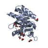
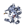
| Deposited unit | 
| |||||||||
|---|---|---|---|---|---|---|---|---|---|---|
| 1 |
| |||||||||
| 2 | 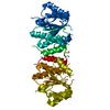
| |||||||||
| Unit cell |
| |||||||||
| Components on special symmetry positions |
|
-Components
| #1: Protein | Mass: 28108.504 Da / Num. of mol.: 1 / Mutation: T242Q Source method: isolated from a genetically manipulated source Source: (gene. exp.) Saccharopolyspora spinosa (bacteria) / Gene: spnH / Plasmid: PET-21B / Production host: Escherichia coli BL21 (bacteria) / Strain (production host): BL21 / References: UniProt: Q9ALM9 |
|---|---|
| #2: Chemical | ChemComp-MG /   Mass: 24.305 Da / Num. of mol.: 1 / Source method: obtained synthetically / Formula: Mg Mass: 24.305 Da / Num. of mol.: 1 / Source method: obtained synthetically / Formula: Mg |
| #3: Water | ChemComp-HOH / Water Mass: 18.015 Da / Num. of mol.: 97 / Source method: isolated from a natural source / Formula: H2O Mass: 18.015 Da / Num. of mol.: 97 / Source method: isolated from a natural source / Formula: H2O |
-Experimental details
-Experiment
| Experiment | Method: X-RAY DIFFRACTION / Number of used crystals: 1 |
|---|
- Sample preparation
| Crystal | Density Matthews: 2.36 Å3/Da / Density % sol: 47.9 % |
|---|---|
| Crystal grow | Temperature: 298 K / Method: vapor diffusion, hanging drop Details: 18-24% (w/v) polyethylene glycol 3350, 100mM Tris, pH 7-9, 200mM magnesium chloride and 2.3mM SRPG PH range: 7-9 |
-Data collection
| Diffraction | Mean temperature: 100 K |
|---|---|
| Diffraction source | Source: SYNCHROTRON / Site: NSRRC / Beamline: BL13B1 / Wavelength: 1 Å |
| Detector | Type: ADSC QUANTUM 270 / Detector: CCD / Date: Nov 28, 2015 / Details: RH COATED MIRROWS |
| Radiation | Monochromator: LN2-COOLED, FIXED-EXIT DOUBLE CRYSTAL MONOCHROMATOR Protocol: SINGLE WAVELENGTH / Monochromatic (M) / Laue (L): M / Scattering type: x-ray |
| Radiation wavelength | Wavelength: 1 Å / Relative weight: 1 |
| Reflection | Resolution: 2→27.6 Å / Num. obs: 26486 / % possible obs: 99.45 % / Observed criterion σ(I): 1 / Redundancy: 38.4 % / Biso Wilson estimate: 34.2 Å2 / Rmerge(I) obs: 0.054 / Net I/σ(I): 39.04 |
| Reflection shell | Resolution: 2.06→2.15 Å / Redundancy: 35.6 % / Rmerge(I) obs: 0.88 / Mean I/σ(I) obs: 3.23 / % possible all: 94.52 |
- Processing
| Software |
| |||||||||||||||||||||||||||||||||||||||||||||||||||||||||||||||||||||||||||||
|---|---|---|---|---|---|---|---|---|---|---|---|---|---|---|---|---|---|---|---|---|---|---|---|---|---|---|---|---|---|---|---|---|---|---|---|---|---|---|---|---|---|---|---|---|---|---|---|---|---|---|---|---|---|---|---|---|---|---|---|---|---|---|---|---|---|---|---|---|---|---|---|---|---|---|---|---|---|---|
| Refinement | Method to determine structure: MOLECULAR REPLACEMENT Starting model: 4CDZ Resolution: 2→27.6 Å / SU ML: 0.28 / Cross valid method: FREE R-VALUE / σ(F): 0 / Phase error: 30.31
| |||||||||||||||||||||||||||||||||||||||||||||||||||||||||||||||||||||||||||||
| Solvent computation | Shrinkage radii: 0.9 Å / VDW probe radii: 1.11 Å | |||||||||||||||||||||||||||||||||||||||||||||||||||||||||||||||||||||||||||||
| Displacement parameters | Biso mean: 46.1 Å2 | |||||||||||||||||||||||||||||||||||||||||||||||||||||||||||||||||||||||||||||
| Refinement step | Cycle: LAST / Resolution: 2→27.6 Å
| |||||||||||||||||||||||||||||||||||||||||||||||||||||||||||||||||||||||||||||
| Refine LS restraints |
| |||||||||||||||||||||||||||||||||||||||||||||||||||||||||||||||||||||||||||||
| LS refinement shell |
|
