 Movie
Movie Controller


+ Open data

- Basic information
| Entry | Database: PDB / ID: 5hxk | |||||||||||||||
|---|---|---|---|---|---|---|---|---|---|---|---|---|---|---|---|---|
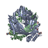
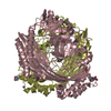
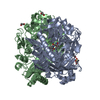
| Title | Structure of TTHA1265 | |||||||||||||||
 Components Components | Zinc-dependent peptidase | |||||||||||||||
 Keywords Keywords |  HYDROLASE / TTHA1265 / protease / complex HYDROLASE / TTHA1265 / protease / complex | |||||||||||||||
| Function / homology | Peptidase M16, C-terminal / Peptidase M16 inactive domain / Peptidase M16, N-terminal / Insulinase (Peptidase family M16) / Metalloenzyme, LuxS/M16 peptidase-like / metal ion binding / Zinc-dependent peptidase Function and homology information Function and homology information | |||||||||||||||
| Biological species |   Thermus thermophilus (bacteria) Thermus thermophilus (bacteria) | |||||||||||||||
| Method | X-RAY DIFFRACTION / SYNCHROTRON / SAD / Resolution: 2 Å | |||||||||||||||
 Authors Authors | Wang, W.W. / Ran, T.T. / Xu, D.Q. / Wang, M.T. | |||||||||||||||
| Funding support |  China, 4items China, 4items
| |||||||||||||||
 Citation Citation | Journal: To Be Published Title: Structure of TTHA1264-1265 complex Authors: Ran, T.T. / Wang, W.W. / Xu, D.Q. / Wang, M.T. | |||||||||||||||
| History |
|
- Structure visualization
| Structure viewer | Molecule: MolmilJmol/JSmol |
|---|
- Downloads & links
-Download
| PDBx/mmCIF format | 5hxk.cif.gz | 252.3 KB | Display | PDBx/mmCIF format |
|---|---|---|---|---|
| PDB format | pdb5hxk.ent.gz | 201.2 KB | Display | PDB format |
| PDBx/mmJSON format | 5hxk.json.gz | Tree view | PDBx/mmJSON format | |
| Others |  Other downloads Other downloads |
-Validation report
| Arichive directory | https://data.pdbj.org/pub/pdb/validation_reports/hx/5hxkftp://data.pdbj.org/pub/pdb/validation_reports/hx/5hxk | HTTPS FTP |
|---|
-Related structure data
| Similar structure data |
|---|

-Links
 PDBj
PDBj

- Assembly
| Deposited unit | 
| ||||||||
|---|---|---|---|---|---|---|---|---|---|
| 1 | 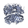
| ||||||||
| 2 | 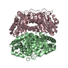
| ||||||||
| Unit cell |
| ||||||||
| Components on special symmetry positions |
|
-Components
| #1: Protein | Mass: 46092.535 Da / Num. of mol.: 3 Source method: isolated from a genetically manipulated source Source: (gene. exp.) Thermus thermophilus (strain HB8 / ATCC 27634 / DSM 579) (bacteria)Strain: HB8 / ATCC 27634 / DSM 579 / Gene: TTHA1265 / Plasmid: pET28a / Production host: Escherichia coli (E. coli) / References: UniProt: Q5SIU9#2: Water | ChemComp-HOH / | Water Mass: 18.015 Da / Num. of mol.: 710 / Source method: isolated from a natural source / Formula: H2O Mass: 18.015 Da / Num. of mol.: 710 / Source method: isolated from a natural source / Formula: H2O |
|---|
-Experimental details
-Experiment
| Experiment | Method: X-RAY DIFFRACTION / Number of used crystals: 1 |
|---|
- Sample preparation
| Crystal | Density Matthews: 2.35 Å3/Da / Density % sol: 47.65 % / Description: rod |
|---|---|
| Crystal grow | Temperature: 295 K / Method: vapor diffusion, sitting drop / pH: 5.5 / Details: PEG 10000, Ammonium acetate |
-Data collection
| Diffraction | Mean temperature: 100 K |
|---|---|
| Diffraction source | Source: SYNCHROTRON / Site: SLS  / Beamline: X06SA / Wavelength: 1 Å / Beamline: X06SA / Wavelength: 1 Å |
| Detector | Type: MARMOSAIC 225 mm CCD / Detector: CCD / Date: Dec 24, 2008 |
| Radiation | Protocol: SINGLE WAVELENGTH / Monochromatic (M) / Laue (L): M / Scattering type: x-ray |
| Radiation wavelength | Wavelength: 1 Å / Relative weight: 1 |
| Reflection | Resolution: 2→20 Å / Num. obs: 88245 / % possible obs: 99.9 % / Redundancy: 7.5 % / Rmerge(I) obs: 0.2 / Net I/σ(I): 8.4 |
| Reflection shell | Resolution: 2→2.11 Å / Redundancy: 7.5 % / Rmerge(I) obs: 0.8 / Mean I/σ(I) obs: 2.6 / % possible all: 100 |
- Processing
| Software |
| |||||||||||||||||||||||||||||||||||||||||||||||||||||||||||||||||||||||||||||||||||||||||||||||||||||||||||||||||||||||||||||||||||||||||||||||||||||||||||||||||||||||||||||||||||||||||||||||||||||||||||||||||||||||||
|---|---|---|---|---|---|---|---|---|---|---|---|---|---|---|---|---|---|---|---|---|---|---|---|---|---|---|---|---|---|---|---|---|---|---|---|---|---|---|---|---|---|---|---|---|---|---|---|---|---|---|---|---|---|---|---|---|---|---|---|---|---|---|---|---|---|---|---|---|---|---|---|---|---|---|---|---|---|---|---|---|---|---|---|---|---|---|---|---|---|---|---|---|---|---|---|---|---|---|---|---|---|---|---|---|---|---|---|---|---|---|---|---|---|---|---|---|---|---|---|---|---|---|---|---|---|---|---|---|---|---|---|---|---|---|---|---|---|---|---|---|---|---|---|---|---|---|---|---|---|---|---|---|---|---|---|---|---|---|---|---|---|---|---|---|---|---|---|---|---|---|---|---|---|---|---|---|---|---|---|---|---|---|---|---|---|---|---|---|---|---|---|---|---|---|---|---|---|---|---|---|---|---|---|---|---|---|---|---|---|---|---|---|---|---|---|---|---|---|
| Refinement | Method to determine structure: SAD / Resolution: 2→19.661 Å / SU ML: 0.23 / Cross valid method: THROUGHOUT / σ(F): 1.34 / Phase error: 22
| |||||||||||||||||||||||||||||||||||||||||||||||||||||||||||||||||||||||||||||||||||||||||||||||||||||||||||||||||||||||||||||||||||||||||||||||||||||||||||||||||||||||||||||||||||||||||||||||||||||||||||||||||||||||||
| Solvent computation | Shrinkage radii: 0.9 Å / VDW probe radii: 1.11 Å | |||||||||||||||||||||||||||||||||||||||||||||||||||||||||||||||||||||||||||||||||||||||||||||||||||||||||||||||||||||||||||||||||||||||||||||||||||||||||||||||||||||||||||||||||||||||||||||||||||||||||||||||||||||||||
| Refinement step | Cycle: LAST / Resolution: 2→19.661 Å
| |||||||||||||||||||||||||||||||||||||||||||||||||||||||||||||||||||||||||||||||||||||||||||||||||||||||||||||||||||||||||||||||||||||||||||||||||||||||||||||||||||||||||||||||||||||||||||||||||||||||||||||||||||||||||
| Refine LS restraints |
| |||||||||||||||||||||||||||||||||||||||||||||||||||||||||||||||||||||||||||||||||||||||||||||||||||||||||||||||||||||||||||||||||||||||||||||||||||||||||||||||||||||||||||||||||||||||||||||||||||||||||||||||||||||||||
| LS refinement shell |
|
