 Movie
Movie Controller

[English] 日本語
 Yorodumi
- PDB-5huv: Structure of Candida albicans trehalose-6-phosphate synthase E341... -
+ Open data

- Basic information
| Entry | Database: PDB / ID: 5huv | ||||||
|---|---|---|---|---|---|---|---|
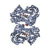
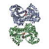
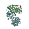
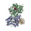
| Title | Structure of Candida albicans trehalose-6-phosphate synthase E341R/E346R in complex with UDP-glucose | ||||||
 Components Components | Alpha,alpha-trehalose-phosphate synthase [UDP-forming] | ||||||
 Keywords Keywords |  TRANSFERASE / trehalose-6-phosphate synthase TRANSFERASE / trehalose-6-phosphate synthase | ||||||
| Function / homology |  Function and homology information Function and homology informationfilamentous growth of a population of unicellular organisms in response to heat / alpha,alpha-trehalose-phosphate synthase complex (UDP-forming) / filamentous growth of a population of unicellular organisms in response to biotic stimulus / cellular response to desiccation / trehalose-phosphatase activity / alpha,alpha-trehalose-phosphate synthase (UDP-forming) / alpha,alpha-trehalose-phosphate synthase (UDP-forming) activity / trehalose biosynthetic process / trehalose metabolism in response to stress / filamentous growth / cellular response to heatSimilarity search - Function | ||||||
| Biological species |  Candida albicans (yeast) Candida albicans (yeast) | ||||||
| Method | X-RAY DIFFRACTION / SYNCHROTRON / MOLECULAR REPLACEMENT / Resolution: 2.002 Å | ||||||
 Authors Authors | Miao, Y. / Brennan, R.G. | ||||||
| Funding support |  United States, 1items United States, 1items
| ||||||
 Citation Citation | Journal: To Be Published Title: Structure of Candida albicans trehalose-6-phosphate synthase E341R/E346R in complex with UDP-glucose Authors: Miao, Y. / Brennan, R.G. | ||||||
| History |
|
- Structure visualization
| Structure viewer | Molecule: MolmilJmol/JSmol |
|---|
- Downloads & links
-Download
| PDBx/mmCIF format | 5huv.cif.gz | 219 KB | Display | PDBx/mmCIF format |
|---|---|---|---|---|
| PDB format | pdb5huv.ent.gz | 172.7 KB | Display | PDB format |
| PDBx/mmJSON format | 5huv.json.gz | Tree view | PDBx/mmJSON format | |
| Others |  Other downloads Other downloads |
-Validation report
| Arichive directory | https://data.pdbj.org/pub/pdb/validation_reports/hu/5huvftp://data.pdbj.org/pub/pdb/validation_reports/hu/5huv | HTTPS FTP |
|---|
-Related structure data
| Related structure data | |
|---|---|
| Similar structure data |


-Links
 PDBj
PDBj
- Assembly
| Deposited unit | 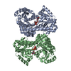
| ||||||||
|---|---|---|---|---|---|---|---|---|---|
| 1 |
| ||||||||
| Unit cell |
| ||||||||
| Components on special symmetry positions |
|
-Components
| #1: Protein | Mass: 54577.336 Da / Num. of mol.: 2 / Mutation: E341R/E346R Source method: isolated from a genetically manipulated source Source: (gene. exp.) Candida albicans (strain SC5314 / ATCC MYA-2876) (yeast)Strain: SC5314 / ATCC MYA-2876 / Gene: TPS1, CaO19.13961, CaO19.6640 / Production host:  Escherichia coli (E. coli) Escherichia coli (E. coli)References: UniProt: Q92410, alpha,alpha-trehalose-phosphate synthase (UDP-forming)#2: Chemical | Uridine diphosphate glucose  Mass: 566.302 Da / Num. of mol.: 2 / Source method: obtained synthetically / Formula: C15H24N2O17P2 Mass: 566.302 Da / Num. of mol.: 2 / Source method: obtained synthetically / Formula: C15H24N2O17P2#3: Water | ChemComp-HOH / | Water Mass: 18.015 Da / Num. of mol.: 735 / Source method: isolated from a natural source / Formula: H2O Mass: 18.015 Da / Num. of mol.: 735 / Source method: isolated from a natural source / Formula: H2O |
|---|
-Experimental details
-Experiment
| Experiment | Method: X-RAY DIFFRACTION / Number of used crystals: 1 |
|---|
- Sample preparation
| Crystal | Density Matthews: 2.4 Å3/Da / Density % sol: 48.76 % |
|---|---|
| Crystal grow | Temperature: 298 K / Method: vapor diffusion, hanging drop / pH: 8.5 / Details: 0.2 M lithium sulfate, 0.1 M Tris, 40% PEG 400 |
-Data collection
| Diffraction | Mean temperature: 100 K | |||||||||||||||||||||||||||||||||||||||||||||||||||||||||||||||||||||||||||||||||||||||||||||||||||||||||||||||||||||||||||||||||||||||||||||||||||||||||||||||||||||||||||||||||||||||||||||
|---|---|---|---|---|---|---|---|---|---|---|---|---|---|---|---|---|---|---|---|---|---|---|---|---|---|---|---|---|---|---|---|---|---|---|---|---|---|---|---|---|---|---|---|---|---|---|---|---|---|---|---|---|---|---|---|---|---|---|---|---|---|---|---|---|---|---|---|---|---|---|---|---|---|---|---|---|---|---|---|---|---|---|---|---|---|---|---|---|---|---|---|---|---|---|---|---|---|---|---|---|---|---|---|---|---|---|---|---|---|---|---|---|---|---|---|---|---|---|---|---|---|---|---|---|---|---|---|---|---|---|---|---|---|---|---|---|---|---|---|---|---|---|---|---|---|---|---|---|---|---|---|---|---|---|---|---|---|---|---|---|---|---|---|---|---|---|---|---|---|---|---|---|---|---|---|---|---|---|---|---|---|---|---|---|---|---|---|---|---|---|
| Diffraction source | Source: SYNCHROTRON / Site: ALS / Beamline: 5.0.1 / Wavelength: 1 Å | |||||||||||||||||||||||||||||||||||||||||||||||||||||||||||||||||||||||||||||||||||||||||||||||||||||||||||||||||||||||||||||||||||||||||||||||||||||||||||||||||||||||||||||||||||||||||||||
| Detector | Type: ADSC QUANTUM 315r / Detector: CCD / Date: Feb 28, 2015 | |||||||||||||||||||||||||||||||||||||||||||||||||||||||||||||||||||||||||||||||||||||||||||||||||||||||||||||||||||||||||||||||||||||||||||||||||||||||||||||||||||||||||||||||||||||||||||||
| Radiation | Protocol: SINGLE WAVELENGTH / Monochromatic (M) / Laue (L): M / Scattering type: x-ray | |||||||||||||||||||||||||||||||||||||||||||||||||||||||||||||||||||||||||||||||||||||||||||||||||||||||||||||||||||||||||||||||||||||||||||||||||||||||||||||||||||||||||||||||||||||||||||||
| Radiation wavelength | Wavelength: 1 Å / Relative weight: 1 | |||||||||||||||||||||||||||||||||||||||||||||||||||||||||||||||||||||||||||||||||||||||||||||||||||||||||||||||||||||||||||||||||||||||||||||||||||||||||||||||||||||||||||||||||||||||||||||
| Reflection twin | Operator: h,-h-k,-l / Fraction: 0.27 | |||||||||||||||||||||||||||||||||||||||||||||||||||||||||||||||||||||||||||||||||||||||||||||||||||||||||||||||||||||||||||||||||||||||||||||||||||||||||||||||||||||||||||||||||||||||||||||
| Reflection | Resolution: 2→50 Å / Num. obs: 69000 / % possible obs: 100 % / Redundancy: 7.5 % / Rmerge(I) obs: 0.091 / Rpim(I) all: 0.036 / Rrim(I) all: 0.098 / Χ2: 1.629 / Net I/av σ(I): 31.5 / Net I/σ(I): 10.3 / Num. measured all: 514978 | |||||||||||||||||||||||||||||||||||||||||||||||||||||||||||||||||||||||||||||||||||||||||||||||||||||||||||||||||||||||||||||||||||||||||||||||||||||||||||||||||||||||||||||||||||||||||||||
| Reflection shell | Diffraction-ID: 1 / Rejects: 0
|
- Processing
| Software |
| ||||||||||||||||||||||||||||||||||||||||||||||||||||||||||||||||||||||||||||||||||||||||||
|---|---|---|---|---|---|---|---|---|---|---|---|---|---|---|---|---|---|---|---|---|---|---|---|---|---|---|---|---|---|---|---|---|---|---|---|---|---|---|---|---|---|---|---|---|---|---|---|---|---|---|---|---|---|---|---|---|---|---|---|---|---|---|---|---|---|---|---|---|---|---|---|---|---|---|---|---|---|---|---|---|---|---|---|---|---|---|---|---|---|---|---|
| Refinement | Method to determine structure: MOLECULAR REPLACEMENT Starting model: Candida albicans Tps1 Resolution: 2.002→49.319 Å / FOM work R set: 0.8481 / Cross valid method: FREE R-VALUE / σ(F): 1.36 / Phase error: 22.12 / Stereochemistry target values: TWIN_LSQ_F
| ||||||||||||||||||||||||||||||||||||||||||||||||||||||||||||||||||||||||||||||||||||||||||
| Solvent computation | Shrinkage radii: 0.9 Å / VDW probe radii: 1.11 Å / Solvent model: FLAT BULK SOLVENT MODEL | ||||||||||||||||||||||||||||||||||||||||||||||||||||||||||||||||||||||||||||||||||||||||||
| Displacement parameters | Biso max: 87.09 Å2 / Biso mean: 26.92 Å2 / Biso min: 10.41 Å2 | ||||||||||||||||||||||||||||||||||||||||||||||||||||||||||||||||||||||||||||||||||||||||||
| Refinement step | Cycle: final / Resolution: 2.002→49.319 Å
| ||||||||||||||||||||||||||||||||||||||||||||||||||||||||||||||||||||||||||||||||||||||||||
| Refine LS restraints |
| ||||||||||||||||||||||||||||||||||||||||||||||||||||||||||||||||||||||||||||||||||||||||||
| LS refinement shell | Refine-ID: X-RAY DIFFRACTION / Total num. of bins used: 14 / % reflection obs: 97 %
|
