 Movie
Movie Controller

[English] 日本語
 Yorodumi
- PDB-5hnk: Crystal structure of T5Fen in complex intact substrate and metal ions. -
+ Open data

- Basic information
| Entry | Database: PDB / ID: 5hnk | ||||||
|---|---|---|---|---|---|---|---|
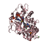
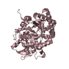
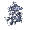
| Title | Crystal structure of T5Fen in complex intact substrate and metal ions. | ||||||
 Components Components |
| ||||||
 Keywords Keywords |  HYDROLASE / enzyme-DNA complex / flap endonuclease / metalloenzyme. HYDROLASE / enzyme-DNA complex / flap endonuclease / metalloenzyme. | ||||||
| Function / homology |  Function and homology informationviral replication complex / exodeoxyribonuclease (lambda-induced) / late viral transcription / DNA replication, Okazaki fragment processing / double-stranded DNA 5'-3' DNA exonuclease activity / DNA exonuclease activity / double-stranded DNA endonuclease activity / 5'-flap endonuclease activity / 5'-3' exonuclease activity / viral DNA genome replication ...viral replication complex / exodeoxyribonuclease (lambda-induced) / late viral transcription / DNA replication, Okazaki fragment processing / double-stranded DNA 5'-3' DNA exonuclease activity / DNA exonuclease activity / double-stranded DNA endonuclease activity / 5'-flap endonuclease activity / 5'-3' exonuclease activity / viral DNA genome replication / Hydrolases; Acting on ester bonds; Exodeoxyribonucleases producing 5'-phosphomonoesters / 5'-3' DNA exonuclease activity / DNA binding / metal ion binding Function and homology informationviral replication complex / exodeoxyribonuclease (lambda-induced) / late viral transcription / DNA replication, Okazaki fragment processing / double-stranded DNA 5'-3' DNA exonuclease activity / DNA exonuclease activity / double-stranded DNA endonuclease activity / 5'-flap endonuclease activity / 5'-3' exonuclease activity / viral DNA genome replication ...viral replication complex / exodeoxyribonuclease (lambda-induced) / late viral transcription / DNA replication, Okazaki fragment processing / double-stranded DNA 5'-3' DNA exonuclease activity / DNA exonuclease activity / double-stranded DNA endonuclease activity / 5'-flap endonuclease activity / 5'-3' exonuclease activity / viral DNA genome replication / Hydrolases; Acting on ester bonds; Exodeoxyribonucleases producing 5'-phosphomonoesters / 5'-3' DNA exonuclease activity / DNA binding / metal ion bindingSimilarity search - Function | ||||||
| Biological species |  Escherichia phage T5 (virus) Escherichia phage T5 (virus)synthetic construct (others) | ||||||
| Method | X-RAY DIFFRACTION / SYNCHROTRON / MOLECULAR REPLACEMENT / Resolution: 2.22 Å | ||||||
 Authors Authors | Almalki, F.A. / Feng, M. / Zhang, J. / Sedelnikova, S.E. / Rafferty, J.B. / Sayers, J.R. / Artymiuk, P.J. | ||||||
 Citation Citation | Journal: Nat.Struct.Mol.Biol. / Year: 2016 Title: Direct observation of DNA threading in flap endonuclease complexes. Authors: AlMalki, F.A. / Flemming, C.S. / Zhang, J. / Feng, M. / Sedelnikova, S.E. / Ceska, T. / Rafferty, J.B. / Sayers, J.R. / Artymiuk, P.J. | ||||||
| History |
|
- Structure visualization
| Structure viewer | Molecule: MolmilJmol/JSmol |
|---|
- Downloads & links
-Download
| PDBx/mmCIF format | 5hnk.cif.gz | 265.4 KB | Display | PDBx/mmCIF format |
|---|---|---|---|---|
| PDB format | pdb5hnk.ent.gz | 210.1 KB | Display | PDB format |
| PDBx/mmJSON format | 5hnk.json.gz | Tree view | PDBx/mmJSON format | |
| Others |  Other downloads Other downloads |
-Validation report
| Arichive directory | https://data.pdbj.org/pub/pdb/validation_reports/hn/5hnkftp://data.pdbj.org/pub/pdb/validation_reports/hn/5hnk | HTTPS FTP |
|---|
-Related structure data
| Related structure data |  5hmlC  5hmmC  5hp4C  1exnS C: citing same article ( S: Starting model for refinement |
|---|---|
| Similar structure data |


-Links
 PDBj
PDBj


- Assembly
| Deposited unit | 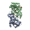
| ||||||||
|---|---|---|---|---|---|---|---|---|---|
| 1 | 
| ||||||||
| 2 | 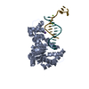
| ||||||||
| Unit cell |
|
-Components
-DNA chain / Protein , 2 types, 4 molecules XYAB
| #1: DNA chain | Mass: 3680.432 Da / Num. of mol.: 2 / Source method: obtained synthetically / Source: (synth.) synthetic construct (others) #2: Protein | / 5'-exonuclease / T5FEN / D15 exonucleaseMass: 31275.475 Da / Num. of mol.: 2 / Fragment: UNP residues 20-291 Source method: isolated from a genetically manipulated source Details: Corresponds to residues 20-291 of Uniprot P06229 but with mutation D153K Source: (gene. exp.) Escherichia phage T5 (virus) / Gene: D15 / Plasmid: pJONEX4Details (production host): pUC19 derivative carrying a lambda promoter. Production host:  Escherichia coli (E. coli) / Strain (production host): M72 Escherichia coli (E. coli) / Strain (production host): M72References: UniProt: P06229, exodeoxyribonuclease (lambda-induced) |
|---|
-Non-polymers , 4 types, 228 molecules 


| #3: Chemical |  Mass: 24.305 Da / Num. of mol.: 2 / Source method: obtained synthetically / Formula: Mg Mass: 24.305 Da / Num. of mol.: 2 / Source method: obtained synthetically / Formula: Mg#4: Chemical | ChemComp-K / |  Mass: 39.098 Da / Num. of mol.: 1 / Source method: obtained synthetically / Formula: K Mass: 39.098 Da / Num. of mol.: 1 / Source method: obtained synthetically / Formula: K#5: Chemical | ChemComp-GOL / | Glycerol Mass: 92.094 Da / Num. of mol.: 1 / Source method: obtained synthetically / Formula: C3H8O3 Mass: 92.094 Da / Num. of mol.: 1 / Source method: obtained synthetically / Formula: C3H8O3#6: Water | ChemComp-HOH / | WaterMass: 18.015 Da / Num. of mol.: 224 / Source method: isolated from a natural source / Formula: H2O |
|---|
-Experimental details
-Experiment
| Experiment | Method: X-RAY DIFFRACTION / Number of used crystals: 1 |
|---|
- Sample preparation
| Crystal | Density Matthews: 2.24 Å3/Da / Density % sol: 45.06 % |
|---|---|
| Crystal grow | Temperature: 290 K / Method: vapor diffusion, hanging drop / pH: 5.5 Details: The concentration of both oligonucleotides was adjusted to 1.1 mM for the duplex molecule by dissolving each one in 10 mM MES pH 6.5 and 50 mM KCl. They were annealed by heating to 367K for ...Details: The concentration of both oligonucleotides was adjusted to 1.1 mM for the duplex molecule by dissolving each one in 10 mM MES pH 6.5 and 50 mM KCl. They were annealed by heating to 367K for 10 minutes and allowed to cool to room temperature. Crystals with oligonucleotide 5ov4 were grown at 290K with the D153K variant of T5Fen. The resulting T5FenD153K:5ov4 structure was determined from crystals grown in 0.2 M MgCl2, 0.1 M Bis-Tris buffer pH 5.5, 25% w/v PEG 3350. |
-Data collection
| Diffraction | Mean temperature: 100 K | ||||||||||||||||||||||||||||||||||||||||||||||||||||||||||||||||||||||||||||||||||||||||||||||||||||||||||||||||||||||||||||||||||||||||||||||||||||||||||||||||||||||||||||||||||||||||||||||||||||||||||||||||||
|---|---|---|---|---|---|---|---|---|---|---|---|---|---|---|---|---|---|---|---|---|---|---|---|---|---|---|---|---|---|---|---|---|---|---|---|---|---|---|---|---|---|---|---|---|---|---|---|---|---|---|---|---|---|---|---|---|---|---|---|---|---|---|---|---|---|---|---|---|---|---|---|---|---|---|---|---|---|---|---|---|---|---|---|---|---|---|---|---|---|---|---|---|---|---|---|---|---|---|---|---|---|---|---|---|---|---|---|---|---|---|---|---|---|---|---|---|---|---|---|---|---|---|---|---|---|---|---|---|---|---|---|---|---|---|---|---|---|---|---|---|---|---|---|---|---|---|---|---|---|---|---|---|---|---|---|---|---|---|---|---|---|---|---|---|---|---|---|---|---|---|---|---|---|---|---|---|---|---|---|---|---|---|---|---|---|---|---|---|---|---|---|---|---|---|---|---|---|---|---|---|---|---|---|---|---|---|---|---|---|---|---|
| Diffraction source | Source: SYNCHROTRON / Site: Diamond  / Beamline: I24 / Wavelength: 0.977 Å / Beamline: I24 / Wavelength: 0.977 Å | ||||||||||||||||||||||||||||||||||||||||||||||||||||||||||||||||||||||||||||||||||||||||||||||||||||||||||||||||||||||||||||||||||||||||||||||||||||||||||||||||||||||||||||||||||||||||||||||||||||||||||||||||||
| Detector | Type: DECTRIS PILATUS 6M / Detector: PIXEL / Date: Apr 16, 2011 Details: Please check which detector was in place at that date | ||||||||||||||||||||||||||||||||||||||||||||||||||||||||||||||||||||||||||||||||||||||||||||||||||||||||||||||||||||||||||||||||||||||||||||||||||||||||||||||||||||||||||||||||||||||||||||||||||||||||||||||||||
| Radiation | Protocol: SINGLE WAVELENGTH / Monochromatic (M) / Laue (L): M / Scattering type: x-ray | ||||||||||||||||||||||||||||||||||||||||||||||||||||||||||||||||||||||||||||||||||||||||||||||||||||||||||||||||||||||||||||||||||||||||||||||||||||||||||||||||||||||||||||||||||||||||||||||||||||||||||||||||||
| Radiation wavelength | Wavelength: 0.977 Å / Relative weight: 1 | ||||||||||||||||||||||||||||||||||||||||||||||||||||||||||||||||||||||||||||||||||||||||||||||||||||||||||||||||||||||||||||||||||||||||||||||||||||||||||||||||||||||||||||||||||||||||||||||||||||||||||||||||||
| Reflection | Resolution: 2.22→44.72 Å / Num. all: 31727 / Num. obs: 31727 / % possible obs: 99.4 % / Redundancy: 5.7 % / Rpim(I) all: 0.038 / Rrim(I) all: 0.094 / Rsym value: 0.079 / Net I/av σ(I): 7.049 / Net I/σ(I): 13.4 / Num. measured all: 182387 | ||||||||||||||||||||||||||||||||||||||||||||||||||||||||||||||||||||||||||||||||||||||||||||||||||||||||||||||||||||||||||||||||||||||||||||||||||||||||||||||||||||||||||||||||||||||||||||||||||||||||||||||||||
| Reflection shell | Diffraction-ID: 1 / Rejects: 0
|
- Processing
| Software |
| |||||||||||||||||||||||||||||||||||||||||||||||||||||||||||||||||||||||||||||||||||||||||||||||||||||||||||||||||||||||||||||
|---|---|---|---|---|---|---|---|---|---|---|---|---|---|---|---|---|---|---|---|---|---|---|---|---|---|---|---|---|---|---|---|---|---|---|---|---|---|---|---|---|---|---|---|---|---|---|---|---|---|---|---|---|---|---|---|---|---|---|---|---|---|---|---|---|---|---|---|---|---|---|---|---|---|---|---|---|---|---|---|---|---|---|---|---|---|---|---|---|---|---|---|---|---|---|---|---|---|---|---|---|---|---|---|---|---|---|---|---|---|---|---|---|---|---|---|---|---|---|---|---|---|---|---|---|---|---|
| Refinement | Method to determine structure: MOLECULAR REPLACEMENT Starting model: 1EXN Resolution: 2.22→42.19 Å / Cor.coef. Fo:Fc: 0.959 / Cor.coef. Fo:Fc free: 0.932 / WRfactor Rfree: 0.2347 / WRfactor Rwork: 0.1834 / FOM work R set: 0.8335 / SU B: 14.251 / SU ML: 0.175 / SU R Cruickshank DPI: 0.3029 / SU Rfree: 0.2192 / Cross valid method: THROUGHOUT / σ(F): 0 / ESU R: 0.303 / ESU R Free: 0.219 / Stereochemistry target values: MAXIMUM LIKELIHOOD Details: HYDROGENS HAVE BEEN ADDED IN THE RIDING POSITIONS U VALUES : WITH TLS ADDED
| |||||||||||||||||||||||||||||||||||||||||||||||||||||||||||||||||||||||||||||||||||||||||||||||||||||||||||||||||||||||||||||
| Solvent computation | Ion probe radii: 0.8 Å / Shrinkage radii: 0.8 Å / VDW probe radii: 1.2 Å / Solvent model: MASK | |||||||||||||||||||||||||||||||||||||||||||||||||||||||||||||||||||||||||||||||||||||||||||||||||||||||||||||||||||||||||||||
| Displacement parameters | Biso max: 154.83 Å2 / Biso mean: 48.335 Å2 / Biso min: 21.96 Å2
| |||||||||||||||||||||||||||||||||||||||||||||||||||||||||||||||||||||||||||||||||||||||||||||||||||||||||||||||||||||||||||||
| Refinement step | Cycle: final / Resolution: 2.22→42.19 Å
| |||||||||||||||||||||||||||||||||||||||||||||||||||||||||||||||||||||||||||||||||||||||||||||||||||||||||||||||||||||||||||||
| Refine LS restraints |
| |||||||||||||||||||||||||||||||||||||||||||||||||||||||||||||||||||||||||||||||||||||||||||||||||||||||||||||||||||||||||||||
| LS refinement shell | Resolution: 2.22→2.278 Å / Total num. of bins used: 20
| |||||||||||||||||||||||||||||||||||||||||||||||||||||||||||||||||||||||||||||||||||||||||||||||||||||||||||||||||||||||||||||
| Refinement TLS params. | Method: refined / Refine-ID: X-RAY DIFFRACTION
| |||||||||||||||||||||||||||||||||||||||||||||||||||||||||||||||||||||||||||||||||||||||||||||||||||||||||||||||||||||||||||||
| Refinement TLS group |
|
