 Movie
Movie Controller

[English] 日本語
 Yorodumi
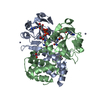
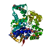
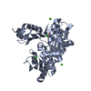
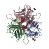
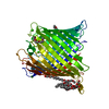
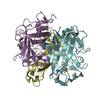
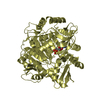
- PDB-5hlj: Crystal Structure of Major Envelope Protein VP24 from White Spot ... -
+ Open data

- Basic information
| Entry | Database: PDB / ID: 5hlj | ||||||
|---|---|---|---|---|---|---|---|
| Title | Crystal Structure of Major Envelope Protein VP24 from White Spot Syndrome Virus | ||||||
 Components Components | VP24 | ||||||
 Keywords Keywords |  VIRAL PROTEIN / WSSV / VP24 / envelope protein VIRAL PROTEIN / WSSV / VP24 / envelope protein | ||||||
| Function / homology | White spot syndrome virus structural envelope protein Vp28 / WSSV envelope protein Vp28 superfamily / White spot syndrome virus structural envelope protein VP / membrane => GO:0016020 / VP24 Function and homology information Function and homology information | ||||||
| Biological species |  White spot syndrome virus White spot syndrome virus | ||||||
| Method | X-RAY DIFFRACTION / SYNCHROTRON / Resolution: 2.406 Å | ||||||
 Authors Authors | Sun, L.F. / Su, Y.T. / Zhao, Y.H. / Fu, Z.Q. / Wu, Y.K. | ||||||
| Funding support |  China, 1items China, 1items
| ||||||
 Citation Citation | Journal: Sci Rep / Year: 2016 Title: Crystal Structure of Major Envelope Protein VP24 from White Spot Syndrome Virus Authors: Sun, L.F. / Su, Y.T. / Zhao, Y.H. / Fu, Z.Q. / Wu, Y.K. | ||||||
| History |
|
- Structure visualization
| Structure viewer | Molecule: MolmilJmol/JSmol |
|---|
- Downloads & links
-Download
| PDBx/mmCIF format | 5hlj.cif.gz | 47.4 KB | Display | PDBx/mmCIF format |
|---|---|---|---|---|
| PDB format | pdb5hlj.ent.gz | 32.9 KB | Display | PDB format |
| PDBx/mmJSON format | 5hlj.json.gz | Tree view | PDBx/mmJSON format | |
| Others |  Other downloads Other downloads |
-Validation report
| Arichive directory | https://data.pdbj.org/pub/pdb/validation_reports/hl/5hljftp://data.pdbj.org/pub/pdb/validation_reports/hl/5hlj | HTTPS FTP |
|---|
-Related structure data
| Similar structure data |
|---|


-Links
 PDBj
PDBj- Assembly
| Deposited unit | 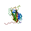
| ||||||||
|---|---|---|---|---|---|---|---|---|---|
| 1 | 
| ||||||||
| Unit cell |
|
-Components
| #1: Protein | Mass: 21255.947 Da / Num. of mol.: 1 / Fragment: UNP residues 30-208 Source method: isolated from a genetically manipulated source Source: (gene. exp.) White spot syndrome virus / Plasmid: pET-21b / Production host:  Escherichia coli BL21(DE3) (bacteria) / Strain (production host): BL21(DE3) / References: UniProt: Q80BF2 Escherichia coli BL21(DE3) (bacteria) / Strain (production host): BL21(DE3) / References: UniProt: Q80BF2 |
|---|---|
| #2: Water | ChemComp-HOH / Water Mass: 18.015 Da / Num. of mol.: 54 / Source method: isolated from a natural source / Formula: H2O Mass: 18.015 Da / Num. of mol.: 54 / Source method: isolated from a natural source / Formula: H2O |
-Experimental details
-Experiment
| Experiment | Method: X-RAY DIFFRACTION / Number of used crystals: 1 |
|---|
- Sample preparation
| Crystal | Density Matthews: 5.35 Å3/Da / Density % sol: 77 % |
|---|---|
| Crystal grow | Temperature: 289 K / Method: vapor diffusion, hanging drop / pH: 8.5 / Details: 0.1M Tris-HCl, pH 8.5, 2.75 M NH4AC |
-Data collection
| Diffraction | Mean temperature: 100 K | |||||||||||||||||||||||||||||||||||||||||||||||||||||||||||||||||||||||||||||||||||||||||||||||||||||||||
|---|---|---|---|---|---|---|---|---|---|---|---|---|---|---|---|---|---|---|---|---|---|---|---|---|---|---|---|---|---|---|---|---|---|---|---|---|---|---|---|---|---|---|---|---|---|---|---|---|---|---|---|---|---|---|---|---|---|---|---|---|---|---|---|---|---|---|---|---|---|---|---|---|---|---|---|---|---|---|---|---|---|---|---|---|---|---|---|---|---|---|---|---|---|---|---|---|---|---|---|---|---|---|---|---|---|---|
| Diffraction source | Source: SYNCHROTRON / Site: SSRF / Beamline: BL17U / Wavelength: 0.97915 Å | |||||||||||||||||||||||||||||||||||||||||||||||||||||||||||||||||||||||||||||||||||||||||||||||||||||||||
| Detector | Type: ADSC QUANTUM 315r / Detector: CCD / Date: May 26, 2015 | |||||||||||||||||||||||||||||||||||||||||||||||||||||||||||||||||||||||||||||||||||||||||||||||||||||||||
| Radiation | Protocol: SINGLE WAVELENGTH / Monochromatic (M) / Laue (L): M / Scattering type: x-ray | |||||||||||||||||||||||||||||||||||||||||||||||||||||||||||||||||||||||||||||||||||||||||||||||||||||||||
| Radiation wavelength | Wavelength: 0.97915 Å / Relative weight: 1 | |||||||||||||||||||||||||||||||||||||||||||||||||||||||||||||||||||||||||||||||||||||||||||||||||||||||||
| Reflection | Resolution: 2.4→100 Å / Num. obs: 17758 / % possible obs: 99.9 % / Redundancy: 20.6 % / Biso Wilson estimate: 71.49 Å2 / Rmerge(I) obs: 0.074 / Net I/av σ(I): 51.361 / Net I/σ(I): 10.5 | |||||||||||||||||||||||||||||||||||||||||||||||||||||||||||||||||||||||||||||||||||||||||||||||||||||||||
| Reflection shell |
|
- Processing
| Software |
| ||||||||||||||||||||||||||||||||||||||||||||||||||||||||||||||||||||||||||||||||||||||||||||||||||
|---|---|---|---|---|---|---|---|---|---|---|---|---|---|---|---|---|---|---|---|---|---|---|---|---|---|---|---|---|---|---|---|---|---|---|---|---|---|---|---|---|---|---|---|---|---|---|---|---|---|---|---|---|---|---|---|---|---|---|---|---|---|---|---|---|---|---|---|---|---|---|---|---|---|---|---|---|---|---|---|---|---|---|---|---|---|---|---|---|---|---|---|---|---|---|---|---|---|---|---|
| Refinement | Starting model: SAD Resolution: 2.406→31.246 Å / SU ML: 0.28 / Cross valid method: FREE R-VALUE / σ(F): 1.34 / Phase error: 22.15 / Stereochemistry target values: ML Details: THE STRUCTURE FACTOR FILE CONTAINS FRIEDEL PAIRS IN F_PLUS/MINUS COLUMNS
| ||||||||||||||||||||||||||||||||||||||||||||||||||||||||||||||||||||||||||||||||||||||||||||||||||
| Solvent computation | Shrinkage radii: 0.9 Å / VDW probe radii: 1.11 Å / Solvent model: FLAT BULK SOLVENT MODEL | ||||||||||||||||||||||||||||||||||||||||||||||||||||||||||||||||||||||||||||||||||||||||||||||||||
| Refinement step | Cycle: LAST / Resolution: 2.406→31.246 Å
| ||||||||||||||||||||||||||||||||||||||||||||||||||||||||||||||||||||||||||||||||||||||||||||||||||
| Refine LS restraints |
| ||||||||||||||||||||||||||||||||||||||||||||||||||||||||||||||||||||||||||||||||||||||||||||||||||
| LS refinement shell |
|
