 Movie
Movie Controller

[English] 日本語
 Yorodumi
- PDB-5hhf: Crystal structure of Aspergillus fumigatus UDP-Galactopyranose mu... -
+ Open data

- Basic information
| Entry | Database: PDB / ID: 5hhf | |||||||||
|---|---|---|---|---|---|---|---|---|---|---|
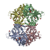
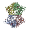
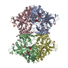
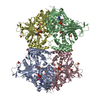
| Title | Crystal structure of Aspergillus fumigatus UDP-Galactopyranose mutase mutant H63A with covalent FAD-Galactopyranose and bound UDP | |||||||||
 Components Components | UDP-galactopyranose mutase | |||||||||
 Keywords Keywords | ISOMERASE / NUCLEOTIDE BINDING / MUTASE / FLAVIN ADENINE DINUCLEOTIDE BINDING / covalent reaction intermediate | |||||||||
| Function / homology |  Function and homology informationUDP-galactopyranose mutase / UDP-galactopyranose mutase activity / nucleotide binding Function and homology informationUDP-galactopyranose mutase / UDP-galactopyranose mutase activity / nucleotide bindingSimilarity search - Function | |||||||||
| Biological species |  Neosartorya fumigata (mold) Neosartorya fumigata (mold) | |||||||||
| Method | X-RAY DIFFRACTION / SYNCHROTRON / FOURIER SYNTHESIS / Resolution: 2.3 Å | |||||||||
 Authors Authors | Tanner, J.J. | |||||||||
| Funding support |  United States, 2items United States, 2items
| |||||||||
 Citation Citation | Journal: Biochemistry / Year: 2016 Title: In Crystallo Capture of a Covalent Intermediate in the UDP-Galactopyranose Mutase Reaction. Authors: Mehra-Chaudhary, R. / Dai, Y. / Sobrado, P. / Tanner, J.J. | |||||||||
| History |
|
- Structure visualization
| Structure viewer | Molecule: MolmilJmol/JSmol |
|---|
- Downloads & links
-Download
| PDBx/mmCIF format | 5hhf.cif.gz | 795 KB | Display | PDBx/mmCIF format |
|---|---|---|---|---|
| PDB format | pdb5hhf.ent.gz | 657.1 KB | Display | PDB format |
| PDBx/mmJSON format | 5hhf.json.gz | Tree view | PDBx/mmJSON format | |
| Others |  Other downloads Other downloads |
-Validation report
| Arichive directory | https://data.pdbj.org/pub/pdb/validation_reports/hh/5hhfftp://data.pdbj.org/pub/pdb/validation_reports/hh/5hhf | HTTPS FTP |
|---|
-Related structure data
| Related structure data | 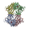 3uthS S: Starting model for refinement |
|---|---|
| Similar structure data |


-Links
 PDBj
PDBj


- Assembly
| Deposited unit | 
| |||||||||||||||
|---|---|---|---|---|---|---|---|---|---|---|---|---|---|---|---|---|
| 1 |
| |||||||||||||||
| Unit cell |
| |||||||||||||||
| Noncrystallographic symmetry (NCS) | NCS domain:
|
-Components
-Protein , 1 types, 4 molecules ABCD
| #1: Protein | Mass: 57061.422 Da / Num. of mol.: 4 / Mutation: H63A Source method: isolated from a genetically manipulated source Source: (gene. exp.) Neosartorya fumigata (mold) / Gene: glf, glfA / Production host:  Escherichia coli (E. coli) / References: UniProt: Q4W1X2, UDP-galactopyranose mutase Escherichia coli (E. coli) / References: UniProt: Q4W1X2, UDP-galactopyranose mutase |
|---|
-Non-polymers , 6 types, 760 molecules 


| #2: Chemical | ChemComp-SO4 / Sulfate Mass: 96.063 Da / Num. of mol.: 10 / Source method: obtained synthetically / Formula: SO4 Mass: 96.063 Da / Num. of mol.: 10 / Source method: obtained synthetically / Formula: SO4#3: Chemical | ChemComp-MRY / Erythritol Mass: 122.120 Da / Num. of mol.: 4 / Source method: obtained synthetically / Formula: C4H10O4 Mass: 122.120 Da / Num. of mol.: 4 / Source method: obtained synthetically / Formula: C4H10O4#4: Chemical |  Mass: 949.706 Da / Num. of mol.: 2 Mass: 949.706 Da / Num. of mol.: 2Source method: isolated from a genetically manipulated source Formula: C33H45N9O20P2 #5: Chemical | Uridine diphosphate Type: RNA linking / Mass: 404.161 Da / Num. of mol.: 2 / Source method: obtained synthetically / Formula: C9H14N2O12P2 / Comment: UDP*YM Type: RNA linking / Mass: 404.161 Da / Num. of mol.: 2 / Source method: obtained synthetically / Formula: C9H14N2O12P2 / Comment: UDP*YM#6: Chemical |  Mass: 787.566 Da / Num. of mol.: 2 / Source method: obtained synthetically / Formula: C27H35N9O15P2 Mass: 787.566 Da / Num. of mol.: 2 / Source method: obtained synthetically / Formula: C27H35N9O15P2#7: Water | ChemComp-HOH / | WaterMass: 18.015 Da / Num. of mol.: 740 / Source method: isolated from a natural source / Formula: H2O |
|---|
-Details
| Sequence details | AUTHOR STATES THAT A429T IS AN UNINTENDED |
|---|
-Experimental details
-Experiment
| Experiment | Method: X-RAY DIFFRACTION / Number of used crystals: 1 |
|---|
- Sample preparation
| Crystal | Density Matthews: 4.8 Å3/Da / Density % sol: 74.37 % |
|---|---|
| Crystal grow | Temperature: 293 K / Method: vapor diffusion, sitting drop / pH: 4.5 Details: 1.6 M ammonium sulfate and 0.1 M sodium acetate at pH 4.5 |
-Data collection
| Diffraction | Mean temperature: 100 K | |||||||||||||||||||||||||||
|---|---|---|---|---|---|---|---|---|---|---|---|---|---|---|---|---|---|---|---|---|---|---|---|---|---|---|---|---|
| Diffraction source | Source: SYNCHROTRON / Site: ALS / Beamline: 4.2.2 / Wavelength: 1 Å | |||||||||||||||||||||||||||
| Detector | Type: RDI CMOS_8M / Detector: CMOS / Date: Sep 4, 2015 | |||||||||||||||||||||||||||
| Radiation | Protocol: SINGLE WAVELENGTH / Monochromatic (M) / Laue (L): M / Scattering type: x-ray | |||||||||||||||||||||||||||
| Radiation wavelength | Wavelength: 1 Å / Relative weight: 1 | |||||||||||||||||||||||||||
| Reflection | Resolution: 2.3→61.61 Å / Num. obs: 196082 / % possible obs: 100 % / Redundancy: 21.1 % / Biso Wilson estimate: 32.33 Å2 / CC1/2: 0.998 / Rmerge(I) obs: 0.201 / Rpim(I) all: 0.045 / Net I/σ(I): 15.1 / Num. measured all: 4132787 | |||||||||||||||||||||||||||
| Reflection shell | Diffraction-ID: 1 / Rejects: 0
|
- Processing
| Software |
| ||||||||||||||||||||||||||||||||||||||||||||||||||||||||||||||||||||||||||||||||||||||||||||||||||||||||||||||||||||||||||||||||||||||||||||||||||||||||||||||||||||||||
|---|---|---|---|---|---|---|---|---|---|---|---|---|---|---|---|---|---|---|---|---|---|---|---|---|---|---|---|---|---|---|---|---|---|---|---|---|---|---|---|---|---|---|---|---|---|---|---|---|---|---|---|---|---|---|---|---|---|---|---|---|---|---|---|---|---|---|---|---|---|---|---|---|---|---|---|---|---|---|---|---|---|---|---|---|---|---|---|---|---|---|---|---|---|---|---|---|---|---|---|---|---|---|---|---|---|---|---|---|---|---|---|---|---|---|---|---|---|---|---|---|---|---|---|---|---|---|---|---|---|---|---|---|---|---|---|---|---|---|---|---|---|---|---|---|---|---|---|---|---|---|---|---|---|---|---|---|---|---|---|---|---|---|---|---|---|---|---|---|---|
| Refinement | Method to determine structure: FOURIER SYNTHESIS Starting model: 3uth Resolution: 2.3→61.61 Å / SU ML: 0.25 / Cross valid method: FREE R-VALUE / σ(F): 1.89 / Phase error: 20.95 / Stereochemistry target values: ML
| ||||||||||||||||||||||||||||||||||||||||||||||||||||||||||||||||||||||||||||||||||||||||||||||||||||||||||||||||||||||||||||||||||||||||||||||||||||||||||||||||||||||||
| Solvent computation | Shrinkage radii: 0.9 Å / VDW probe radii: 1.11 Å / Solvent model: FLAT BULK SOLVENT MODEL | ||||||||||||||||||||||||||||||||||||||||||||||||||||||||||||||||||||||||||||||||||||||||||||||||||||||||||||||||||||||||||||||||||||||||||||||||||||||||||||||||||||||||
| Displacement parameters | Biso max: 127.55 Å2 / Biso mean: 39.5653 Å2 / Biso min: 18.74 Å2 | ||||||||||||||||||||||||||||||||||||||||||||||||||||||||||||||||||||||||||||||||||||||||||||||||||||||||||||||||||||||||||||||||||||||||||||||||||||||||||||||||||||||||
| Refinement step | Cycle: final / Resolution: 2.3→61.61 Å
| ||||||||||||||||||||||||||||||||||||||||||||||||||||||||||||||||||||||||||||||||||||||||||||||||||||||||||||||||||||||||||||||||||||||||||||||||||||||||||||||||||||||||
| Refine LS restraints |
| ||||||||||||||||||||||||||||||||||||||||||||||||||||||||||||||||||||||||||||||||||||||||||||||||||||||||||||||||||||||||||||||||||||||||||||||||||||||||||||||||||||||||
| Refine LS restraints NCS |
| ||||||||||||||||||||||||||||||||||||||||||||||||||||||||||||||||||||||||||||||||||||||||||||||||||||||||||||||||||||||||||||||||||||||||||||||||||||||||||||||||||||||||
| LS refinement shell | Refine-ID: X-RAY DIFFRACTION / Total num. of bins used: 27 / % reflection obs: 100 %
| ||||||||||||||||||||||||||||||||||||||||||||||||||||||||||||||||||||||||||||||||||||||||||||||||||||||||||||||||||||||||||||||||||||||||||||||||||||||||||||||||||||||||
| Refinement TLS params. | Method: refined / Refine-ID: X-RAY DIFFRACTION
| ||||||||||||||||||||||||||||||||||||||||||||||||||||||||||||||||||||||||||||||||||||||||||||||||||||||||||||||||||||||||||||||||||||||||||||||||||||||||||||||||||||||||
| Refinement TLS group |
|
