Monochromator: double crystal / Protocol: SINGLE WAVELENGTH / Monochromatic (M) / Laue (L): M / Scattering type: x-ray
Radiation wavelength
Wavelength: 1.2827 Å / Relative weight: 1
Reflection
Redundancy: 7.2 % / Number: 184019 / Rmerge(I) obs: 0.092 / Χ2: 3.31 / D res high: 1.9 Å / D res low: 50 Å / Num. obs: 25599 / % possible obs: 99.8
Diffraction reflection shell
Highest resolution (Å)
Lowest resolution (Å)
ID
Rmerge(I) obs
Chi squared
Redundancy
4.09
50
1
0.071
8.067
6.6
3.25
4.09
1
0.075
6.04
6.7
2.84
3.25
1
0.089
4.802
7
2.58
2.84
1
0.107
3.426
7.3
2.39
2.58
1
0.127
2.81
7.4
2.25
2.39
1
0.157
2.328
7.4
2.14
2.25
1
0.196
2.029
7.4
2.05
2.14
1
0.251
1.684
7.4
1.97
2.05
1
0.345
1.437
7.4
1.9
1.97
1
0.491
1.298
7.4
Reflection
Resolution: 1.9→50 Å / Num. obs: 25599 / % possible obs: 99.8 % / Redundancy: 7.2 % / Rmerge(I) obs: 0.092 / Χ2: 3.306 / Net I/av σ(I): 46.324 / Net I/σ(I): 14 / Num. measured all: 184019
Reflection shell
Diffraction-ID: 1 / Rejects: 0
Resolution (Å)
Redundancy (%)
Rmerge(I) obs
Num. unique all
Χ2
% possible all
1.9-1.97
7.4
0.491
2524
1.298
99.6
1.97-2.05
7.4
0.345
2538
1.437
99.5
2.05-2.14
7.4
0.251
2551
1.684
99.8
2.14-2.25
7.4
0.196
2553
2.029
99.8
2.25-2.39
7.4
0.157
2543
2.328
99.8
2.39-2.58
7.4
0.127
2570
2.81
99.9
2.58-2.84
7.3
0.107
2557
3.426
100
2.84-3.25
7
0.089
2563
4.802
100
3.25-4.09
6.7
0.075
2581
6.04
100
4.09-50
6.6
0.071
2619
8.067
99.6
-
Phasing
Phasing
Method: SAD
-
Processing
Software
Name
Version
Classification
HKL-2000
datascaling
PHENIX
1.7.3_928
refinement
PDB_EXTRACT
3.15
dataextraction
HKL-2000
datareduction
PHENIX
phasing
Refinement
Method to determine structure: SAD / Resolution: 1.901→45.543 Å / FOM work R set: 0.8747 / SU ML: 0.24 / Cross valid method: FREE R-VALUE / σ(F): 1.22 / Phase error: 20.1 / Stereochemistry target values: ML Details: ANOMALOUS DATA WAS USED FOR REFINEMENT. SF FILE CONTAINS FRIEDEL PAIRS UNDER I/F_MINUS AND I/F_PLUS COLUMNS.
Rfactor
Num. reflection
% reflection
Rfree
0.2047
2565
5.13 %
Rwork
0.1943
47421
-
obs
0.1948
25576
99.27 %
Solvent computation
Shrinkage radii: 0.86 Å / VDW probe radii: 1.1 Å / Solvent model: FLAT BULK SOLVENT MODEL / Bsol: 36.682 Å2 / ksol: 0.345 e/Å3
In the structure databanks used in Yorodumi, some data are registered as the other names, "COVID-19 virus" and "2019-nCoV". Here are the details of the virus and the list of structure data.
Jan 31, 2019. EMDB accession codes are about to change! (news from PDBe EMDB page)
EMDB accession codes are about to change! (news from PDBe EMDB page)
The allocation of 4 digits for EMDB accession codes will soon come to an end. Whilst these codes will remain in use, new EMDB accession codes will include an additional digit and will expand incrementally as the available range of codes is exhausted. The current 4-digit format prefixed with “EMD-” (i.e. EMD-XXXX) will advance to a 5-digit format (i.e. EMD-XXXXX), and so on. It is currently estimated that the 4-digit codes will be depleted around Spring 2019, at which point the 5-digit format will come into force.
The EM Navigator/Yorodumi systems omit the EMD- prefix.
Related info.:Q: What is EMD? / ID/Accession-code notation in Yorodumi/EM Navigator
Yorodumi is a browser for structure data from EMDB, PDB, SASBDB, etc.
This page is also the successor to EM Navigator detail page, and also detail information page/front-end page for Omokage search.
The word "yorodu" (or yorozu) is an old Japanese word meaning "ten thousand". "mi" (miru) is to see.
Related info.:EMDB / PDB / SASBDB / Comparison of 3 databanks / Yorodumi Search / Aug 31, 2016. New EM Navigator & Yorodumi / Yorodumi Papers / Jmol/JSmol / Function and homology information / Changes in new EM Navigator and Yorodumi
 Movie
Movie Controller

 Yorodumi
 Open data
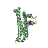
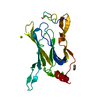
 Basic information
 Components
 Keywords
 TRANSCRIPTION /
  Function and homology information
 Authors
 Citation
 Structure visualization
 Downloads & links
 Other downloads


 PDBj
PDBj


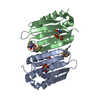
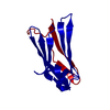
 Assembly


 Mass: 65.409 Da / Num. of mol.: 2 / Source method: obtained synthetically / Formula: Zn
 Mass: 18.015 Da / Num. of mol.: 189 / Source method: isolated from a natural source / Formula: H2O
 Sample preparation
 / Beamline: BL17U / Wavelength: 1.2827 Å
 Processing