 Movie
Movie Controller


+ Open data

- Basic information
| Entry | Database: PDB / ID: 5hh0 | ||||||||||||
|---|---|---|---|---|---|---|---|---|---|---|---|---|---|
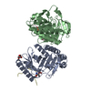
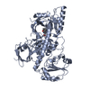
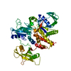
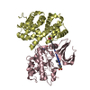
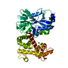
| Title | Crystal structure of human Naa60 in complex with CoA | ||||||||||||
 Components Components | N-alpha-acetyltransferase 60 | ||||||||||||
 Keywords Keywords |  TRANSFERASE / N-terminal acetylation / NATs / Protein modification TRANSFERASE / N-terminal acetylation / NATs / Protein modification | ||||||||||||
| Function / homology |  Function and homology information Function and homology informationN-terminal methionine Nalpha-acetyltransferase NatF / N-terminal peptidyl-methionine acetylation / N-terminal protein amino acid acetylation / peptide alpha-N-acetyltransferase activity / histone H4 acetyltransferase activity / histone acetyltransferase activity / histone acetyltransferase / chromosome segregation / nucleosome assembly / cell population proliferation ...N-terminal methionine Nalpha-acetyltransferase NatF / N-terminal peptidyl-methionine acetylation / N-terminal protein amino acid acetylation / peptide alpha-N-acetyltransferase activity / histone H4 acetyltransferase activity / histone acetyltransferase activity / histone acetyltransferase / chromosome segregation / nucleosome assembly / cell population proliferation / Golgi membrane / protein homodimerization activitySimilarity search - Function | ||||||||||||
| Biological species |  Homo sapiens (human) Homo sapiens (human) | ||||||||||||
| Method | X-RAY DIFFRACTION / SYNCHROTRON / MOLECULAR REPLACEMENT / Resolution: 1.6 Å | ||||||||||||
 Authors Authors | Chen, J.Y. / Liu, L. / Yun, C.H. | ||||||||||||
| Funding support |  China, 3items China, 3items
| ||||||||||||
 Citation Citation | Journal: Sci Rep / Year: 2016 Title: Structure and function of human Naa60 (NatF), a Golgi-localized bi-functional acetyltransferase Authors: Chen, J.Y. / Liu, L. / Cao, C.L. / Li, M.J. / Tan, K. / Yang, X. / Yun, C.H. | ||||||||||||
| History |
|
- Structure visualization
| Structure viewer | Molecule: MolmilJmol/JSmol |
|---|
- Downloads & links
-Download
| PDBx/mmCIF format | 5hh0.cif.gz | 62.9 KB | Display | PDBx/mmCIF format |
|---|---|---|---|---|
| PDB format | pdb5hh0.ent.gz | 44 KB | Display | PDB format |
| PDBx/mmJSON format | 5hh0.json.gz | Tree view | PDBx/mmJSON format | |
| Others |  Other downloads Other downloads |
-Validation report
| Arichive directory | https://data.pdbj.org/pub/pdb/validation_reports/hh/5hh0ftp://data.pdbj.org/pub/pdb/validation_reports/hh/5hh0 | HTTPS FTP |
|---|
-Related structure data
| Related structure data |  5hgzSC 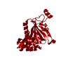 5hh1C C: citing same article ( S: Starting model for refinement |
|---|---|
| Similar structure data |


-Links
 PDBj
PDBj


- Assembly
| Deposited unit | 
| ||||||||
|---|---|---|---|---|---|---|---|---|---|
| 1 | 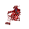
| ||||||||
| Unit cell |
|
-Components
| #1: Protein | Mass: 22968.012 Da / Num. of mol.: 1 / Fragment: UNP residues 1-199 / Mutation: V4E, V5E, P6R Source method: isolated from a genetically manipulated source Source: (gene. exp.) Homo sapiens (human) / Gene: NAA60, HAT4, NAT15, UNQ2771/PRO7155 / Production host:  Escherichia coli (E. coli) Escherichia coli (E. coli)References: UniProt: Q9H7X0, histone acetyltransferase, EC: 2.3.1.88 |
|---|---|
| #2: Chemical | ChemComp-COA / Coenzyme A  Mass: 767.534 Da / Num. of mol.: 1 / Source method: obtained synthetically / Formula: C21H36N7O16P3S Mass: 767.534 Da / Num. of mol.: 1 / Source method: obtained synthetically / Formula: C21H36N7O16P3S |
| #3: Water | ChemComp-HOH / Water Mass: 18.015 Da / Num. of mol.: 258 / Source method: isolated from a natural source / Formula: H2O Mass: 18.015 Da / Num. of mol.: 258 / Source method: isolated from a natural source / Formula: H2O |
-Experimental details
-Experiment
| Experiment | Method: X-RAY DIFFRACTION / Number of used crystals: 1 |
|---|
- Sample preparation
| Crystal | Density Matthews: 2.32 Å3/Da / Density % sol: 46.87 % |
|---|---|
| Crystal grow | Temperature: 293.15 K / Method: liquid diffusion / pH: 7.5 Details: 0.2 M L-Proline, 0.1 M HEPES pH7.5, 10% w/v PEG 3350 |
-Data collection
| Diffraction | Mean temperature: 100 K |
|---|---|
| Diffraction source | Source: SYNCHROTRON / Site: APS  / Beamline: 19-ID / Wavelength: 0.97934 Å / Beamline: 19-ID / Wavelength: 0.97934 Å |
| Detector | Type: ADSC QUANTUM 315r / Detector: CCD / Date: Apr 15, 2015 |
| Radiation | Protocol: SINGLE WAVELENGTH / Monochromatic (M) / Laue (L): M / Scattering type: x-ray |
| Radiation wavelength | Wavelength: 0.97934 Å / Relative weight: 1 |
| Reflection | Resolution: 1.6→50 Å / Num. obs: 28597 / % possible obs: 99.6 % / Redundancy: 6.9 % / Rmerge(I) obs: 0.021 / Net I/σ(I): 31.8 |
| Reflection shell | Resolution: 1.6→1.66 Å / Redundancy: 6.2 % / Rmerge(I) obs: 0.325 / Mean I/σ(I) obs: 2 / % possible all: 98.5 |
- Processing
| Software |
| |||||||||||||||||||||||||||||||||||||||||||||||||||||||||||||||||||||||||||||
|---|---|---|---|---|---|---|---|---|---|---|---|---|---|---|---|---|---|---|---|---|---|---|---|---|---|---|---|---|---|---|---|---|---|---|---|---|---|---|---|---|---|---|---|---|---|---|---|---|---|---|---|---|---|---|---|---|---|---|---|---|---|---|---|---|---|---|---|---|---|---|---|---|---|---|---|---|---|---|
| Refinement | Method to determine structure: MOLECULAR REPLACEMENT Starting model: 5HGZ Resolution: 1.6→33.55 Å / SU ML: 0.18 / Cross valid method: FREE R-VALUE / σ(F): 1.36 / Phase error: 22.65 / Stereochemistry target values: ML
| |||||||||||||||||||||||||||||||||||||||||||||||||||||||||||||||||||||||||||||
| Solvent computation | Shrinkage radii: 0.9 Å / VDW probe radii: 1.11 Å / Solvent model: FLAT BULK SOLVENT MODEL | |||||||||||||||||||||||||||||||||||||||||||||||||||||||||||||||||||||||||||||
| Refinement step | Cycle: LAST / Resolution: 1.6→33.55 Å
| |||||||||||||||||||||||||||||||||||||||||||||||||||||||||||||||||||||||||||||
| Refine LS restraints |
| |||||||||||||||||||||||||||||||||||||||||||||||||||||||||||||||||||||||||||||
| LS refinement shell |
|
