 Movie
Movie Controller

[English] 日本語
 Yorodumi
- PDB-5hcv: Identification of Spirooxindole and Dibenzoxazepine Motifs as Pot... -
+ Open data

- Basic information
| Entry | Database: PDB / ID: 5hcv | ||||||
|---|---|---|---|---|---|---|---|
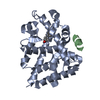
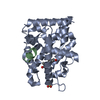
| Title | Identification of Spirooxindole and Dibenzoxazepine Motifs as Potent Mineralocorticoid Receptor Antagonists | ||||||
 Components Components | Mineralocorticoid receptor | ||||||
 Keywords Keywords | SIGNALING PROTEIN / Mineralocorticoid Receptor / Ligand-binding domain / MR-LBD / Antagonists / co-crystal | ||||||
| Function / homology |  Function and homology information Function and homology informationnuclear steroid receptor activity / estrogen response element binding / intracellular steroid hormone receptor signaling pathway / HSP90 chaperone cycle for steroid hormone receptors (SHR) in the presence of ligand / TBP-class protein binding / steroid binding / SUMOylation of intracellular receptors / Nuclear Receptor transcription pathway / positive regulation of non-canonical NF-kappaB signal transduction / nuclear receptor activity ...nuclear steroid receptor activity / estrogen response element binding / intracellular steroid hormone receptor signaling pathway / HSP90 chaperone cycle for steroid hormone receptors (SHR) in the presence of ligand / TBP-class protein binding / steroid binding / SUMOylation of intracellular receptors / Nuclear Receptor transcription pathway / positive regulation of non-canonical NF-kappaB signal transduction / nuclear receptor activity / sequence-specific double-stranded DNA binding / receptor complex / DNA-binding transcription factor activity, RNA polymerase II-specific / DNA-binding transcription factor activity / chromatin / endoplasmic reticulum membrane / regulation of transcription by RNA polymerase II / signal transduction / zinc ion binding / nucleoplasm / cytosolSimilarity search - Function | ||||||
| Biological species |  Homo sapiens (human) Homo sapiens (human) | ||||||
| Method | X-RAY DIFFRACTION / SYNCHROTRON / MOLECULAR REPLACEMENT / Resolution: 2.5 Å | ||||||
 Authors Authors | Chen, G. / McKeever, B.M. | ||||||
 Citation Citation | Journal: Bioorg.Med.Chem. / Year: 2016 Title: Identification of spirooxindole and dibenzoxazepine motifs as potent mineralocorticoid receptor antagonists. Authors: Lotesta, S.D. / Marcus, A.P. / Zheng, Y. / Leftheris, K. / Noto, P.B. / Meng, S. / Kandpal, G. / Chen, G. / Zhou, J. / McKeever, B. / Bukhtiyarov, Y. / Zhao, Y. / Lala, D.S. / Singh, S.B. / McGeehan, G.M. | ||||||
| History |
|
- Structure visualization
| Structure viewer | Molecule: MolmilJmol/JSmol |
|---|
- Downloads & links
-Download
| PDBx/mmCIF format | 5hcv.cif.gz | 163.1 KB | Display | PDBx/mmCIF format |
|---|---|---|---|---|
| PDB format | pdb5hcv.ent.gz | 127.1 KB | Display | PDB format |
| PDBx/mmJSON format | 5hcv.json.gz | Tree view | PDBx/mmJSON format | |
| Others |  Other downloads Other downloads |
-Validation report
| Arichive directory | https://data.pdbj.org/pub/pdb/validation_reports/hc/5hcvftp://data.pdbj.org/pub/pdb/validation_reports/hc/5hcv | HTTPS FTP |
|---|
-Related structure data
| Related structure data |  2a3iS S: Starting model for refinement |
|---|---|
| Similar structure data |

-Links
 PDBj
PDBj

- Assembly
| Deposited unit | 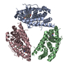
| |||||||||||||||||||||||||||||||||||||||||||||||||||||
|---|---|---|---|---|---|---|---|---|---|---|---|---|---|---|---|---|---|---|---|---|---|---|---|---|---|---|---|---|---|---|---|---|---|---|---|---|---|---|---|---|---|---|---|---|---|---|---|---|---|---|---|---|---|---|
| 1 | 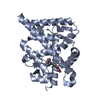
| |||||||||||||||||||||||||||||||||||||||||||||||||||||
| 2 | 
| |||||||||||||||||||||||||||||||||||||||||||||||||||||
| 3 | 
| |||||||||||||||||||||||||||||||||||||||||||||||||||||
| Unit cell |
| |||||||||||||||||||||||||||||||||||||||||||||||||||||
| Noncrystallographic symmetry (NCS) | NCS domain:
NCS domain segments: Component-ID: 0 / Beg auth comp-ID: PRO / Beg label comp-ID: PRO / End auth comp-ID: HIS / End label comp-ID: HIS / Refine code: 0 / Auth seq-ID: 738 - 982 / Label seq-ID: 11 - 255
NCS ensembles :
|
-Components
| #1: Protein | / MR / Nuclear receptor subfamily 3 group C member 2 Mass: 29873.594 Da / Num. of mol.: 3 / Fragment: ligand-binding domain (UNP residues 732-984) / Mutation: C808S Source method: isolated from a genetically manipulated source Source: (gene. exp.) Homo sapiens (human) / Gene: NR3C2, MCR, MLR / Plasmid: pMAL_c5x / Production host:  Escherichia coli (E. coli) / Strain (production host): BL21 (DE3) / References: UniProt: P08235 Escherichia coli (E. coli) / Strain (production host): BL21 (DE3) / References: UniProt: P08235#2: Chemical |   Mass: 373.376 Da / Num. of mol.: 3 / Source method: obtained synthetically / Formula: C23H16FNO3 Mass: 373.376 Da / Num. of mol.: 3 / Source method: obtained synthetically / Formula: C23H16FNO3#3: Chemical | ChemComp-CL / | Chloride  Mass: 35.453 Da / Num. of mol.: 1 / Source method: obtained synthetically / Formula: Cl Mass: 35.453 Da / Num. of mol.: 1 / Source method: obtained synthetically / Formula: Cl#4: Water | ChemComp-HOH / | Water Mass: 18.015 Da / Num. of mol.: 260 / Source method: isolated from a natural source / Formula: H2O Mass: 18.015 Da / Num. of mol.: 260 / Source method: isolated from a natural source / Formula: H2O |
|---|
-Experimental details
-Experiment
| Experiment | Method: X-RAY DIFFRACTION |
|---|
- Sample preparation
| Crystal | Density Matthews: 2.22 Å3/Da / Density % sol: 44.53 % |
|---|---|
| Crystal grow | Temperature: 294 K / Method: vapor diffusion, hanging drop / pH: 8 Details: 1uL of protein complex at 8-10mg protein/ml is mixed with an equal volume of reservoir solution composed of 100mM Tris-HCl pH=8.0, 200mM NaCl, 5% (v/v) glycerol, 16%(w/v) PEG3350 PH range: 7.5 - 8.5 |
-Data collection
| Diffraction | Mean temperature: 100 K / Ambient temp details: CryoJet 7000 |
|---|---|
| Diffraction source | Source: SYNCHROTRON / Site: NSLS  / Beamline: X29A / Wavelength: 1.075 Å / Beamline: X29A / Wavelength: 1.075 Å |
| Detector | Type: ADSC QUANTUM 315r / Detector: CCD / Date: Apr 28, 2011 Details: Cryogenically-cooled monochromator+vertical focusing mirror |
| Radiation | Monochromator: sagittally-focused monochromator / Protocol: SINGLE WAVELENGTH / Monochromatic (M) / Laue (L): M / Scattering type: x-ray |
| Radiation wavelength | Wavelength: 1.075 Å / Relative weight: 1 |
| Reflection | Resolution: 2.5→44.41 Å / Num. all: 25409 / Num. obs: 24098 / % possible obs: 99.97 % / Observed criterion σ(I): 1 / Redundancy: 10.9 % / Rmerge(I) obs: 0.165 / Net I/σ(I): 18.5 |
| Reflection shell | Resolution: 2.499→2.564 Å / Redundancy: 8.3 % / Rmerge(I) obs: 0.95 / Mean I/σ(I) obs: 2.6 / % possible all: 99.79 |
- Processing
| Software |
| ||||||||||||||||||||||||||||||||||||||||||||||||||||||||||||||||||||||||||||||||||||||||||||||||||||||||||||||||||||||||||||||||||||||||||||||||||||||||||||||||||||||||||||||||||||||
|---|---|---|---|---|---|---|---|---|---|---|---|---|---|---|---|---|---|---|---|---|---|---|---|---|---|---|---|---|---|---|---|---|---|---|---|---|---|---|---|---|---|---|---|---|---|---|---|---|---|---|---|---|---|---|---|---|---|---|---|---|---|---|---|---|---|---|---|---|---|---|---|---|---|---|---|---|---|---|---|---|---|---|---|---|---|---|---|---|---|---|---|---|---|---|---|---|---|---|---|---|---|---|---|---|---|---|---|---|---|---|---|---|---|---|---|---|---|---|---|---|---|---|---|---|---|---|---|---|---|---|---|---|---|---|---|---|---|---|---|---|---|---|---|---|---|---|---|---|---|---|---|---|---|---|---|---|---|---|---|---|---|---|---|---|---|---|---|---|---|---|---|---|---|---|---|---|---|---|---|---|---|---|---|
| Refinement | Method to determine structure: MOLECULAR REPLACEMENT Starting model: 2A3I Resolution: 2.5→44.41 Å / Cor.coef. Fo:Fc: 0.958 / Cor.coef. Fo:Fc free: 0.895 / SU B: 11.238 / SU ML: 0.249 / Cross valid method: THROUGHOUT / ESU R Free: 0.327 / Stereochemistry target values: MAXIMUM LIKELIHOOD
| ||||||||||||||||||||||||||||||||||||||||||||||||||||||||||||||||||||||||||||||||||||||||||||||||||||||||||||||||||||||||||||||||||||||||||||||||||||||||||||||||||||||||||||||||||||||
| Solvent computation | Ion probe radii: 0.8 Å / Shrinkage radii: 0.8 Å / VDW probe radii: 1.2 Å / Solvent model: BABINET MODEL WITH MASK | ||||||||||||||||||||||||||||||||||||||||||||||||||||||||||||||||||||||||||||||||||||||||||||||||||||||||||||||||||||||||||||||||||||||||||||||||||||||||||||||||||||||||||||||||||||||
| Displacement parameters | Biso mean: 50.713 Å2
| ||||||||||||||||||||||||||||||||||||||||||||||||||||||||||||||||||||||||||||||||||||||||||||||||||||||||||||||||||||||||||||||||||||||||||||||||||||||||||||||||||||||||||||||||||||||
| Refinement step | Cycle: LAST / Resolution: 2.5→44.41 Å
| ||||||||||||||||||||||||||||||||||||||||||||||||||||||||||||||||||||||||||||||||||||||||||||||||||||||||||||||||||||||||||||||||||||||||||||||||||||||||||||||||||||||||||||||||||||||
| Refine LS restraints |
|