 Movie
Movie Controller


+ Open data

- Basic information
| Entry | Database: PDB / ID: 5h87 | ||||||
|---|---|---|---|---|---|---|---|
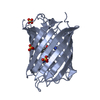
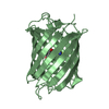
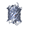
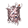
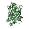
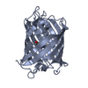
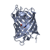
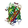
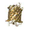
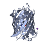
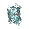
| Title | Crystal structure of mRojoA mutant - P63H - W143S | ||||||
 Components Components | mRojoA fluorescent protein | ||||||
 Keywords Keywords |  FLUORESCENT PROTEIN / quantum yield / protein engineering FLUORESCENT PROTEIN / quantum yield / protein engineering | ||||||
| Function / homology |  Function and homology information Function and homology information | ||||||
| Biological species |  Discosoma sp. (sea anemone) Discosoma sp. (sea anemone) | ||||||
| Method | X-RAY DIFFRACTION / SYNCHROTRON / Resolution: 2.24 Å | ||||||
 Authors Authors | Pandelieva, A.T. / Tremblay, V. / Sarvan, S. / Chica, R.A. / Couture, J.-F. | ||||||
| Funding support |  Canada, 1items Canada, 1items
| ||||||
 Citation Citation | Journal: Acs Chem.Biol. / Year: 2016 Title: Brighter Red Fluorescent Proteins by Rational Design of Triple-Decker Motif. Authors: Pandelieva, A.T. / Baran, M.J. / Calderini, G.F. / McCann, J.L. / Tremblay, V. / Sarvan, S. / Davey, J.A. / Couture, J.F. / Chica, R.A. | ||||||
| History |
|
- Structure visualization
| Structure viewer | Molecule: MolmilJmol/JSmol |
|---|
- Downloads & links
-Download
| PDBx/mmCIF format | 5h87.cif.gz | 181.4 KB | Display | PDBx/mmCIF format |
|---|---|---|---|---|
| PDB format | pdb5h87.ent.gz | 151.7 KB | Display | PDB format |
| PDBx/mmJSON format | 5h87.json.gz | Tree view | PDBx/mmJSON format | |
| Others |  Other downloads Other downloads |
-Validation report
| Arichive directory | https://data.pdbj.org/pub/pdb/validation_reports/h8/5h87ftp://data.pdbj.org/pub/pdb/validation_reports/h8/5h87 | HTTPS FTP |
|---|
-Related structure data


-Links
 PDBj
PDBj

- Assembly
| Deposited unit | 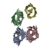
| ||||||||
|---|---|---|---|---|---|---|---|---|---|
| 1 | 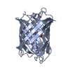
| ||||||||
| 2 | 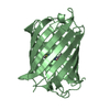
| ||||||||
| 3 | 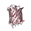
| ||||||||
| 4 | 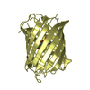
| ||||||||
| Unit cell |
|
-Components
| #1: Protein | Mass: 27647.049 Da / Num. of mol.: 4 / Mutation: P63H and W143S Source method: isolated from a genetically manipulated source Source: (gene. exp.) Discosoma sp. (sea anemone) / Gene: PAmCherry / Production host:  Escherichia coli (E. coli) / References: UniProt: D1MPT3*PLUS Escherichia coli (E. coli) / References: UniProt: D1MPT3*PLUS#2: Water | ChemComp-HOH / | Water Mass: 18.015 Da / Num. of mol.: 217 / Source method: isolated from a natural source / Formula: H2O Mass: 18.015 Da / Num. of mol.: 217 / Source method: isolated from a natural source / Formula: H2OSource details | THIS SEQUENCE WAS BASED ON THE PAMCHERRY1 PROTEIN (UNP REFERENCE D1MPT3). RESIDUES MET 64, TYR 65, ...THIS SEQUENCE WAS BASED ON THE PAMCHERRY1 PROTEIN (UNP REFERENCE D1MPT3). RESIDUES MET 64, TYR 65, AND GLY 68 form a CHROMOPHOR | |
|---|
-Experimental details
-Experiment
| Experiment | Method: X-RAY DIFFRACTION |
|---|
- Sample preparation
| Crystal | Density Matthews: 2.06 Å3/Da / Density % sol: 40.22 % |
|---|---|
| Crystal grow | Temperature: 298 K / Method: vapor diffusion, sitting drop / pH: 5.5 / Details: 125mM Bis-Tris, and 25% Peg3350 |
-Data collection
| Diffraction | Mean temperature: 100 K |
|---|---|
| Diffraction source | Source: SYNCHROTRON / Site: APS  / Beamline: 17-ID / Wavelength: 1 Å / Beamline: 17-ID / Wavelength: 1 Å |
| Detector | Type: MARMOSAIC 225 mm CCD / Detector: CCD / Date: Dec 11, 2014 |
| Radiation | Protocol: SINGLE WAVELENGTH / Monochromatic (M) / Laue (L): M / Scattering type: x-ray |
| Radiation wavelength | Wavelength: 1 Å / Relative weight: 1 |
| Reflection | Resolution: 2.24→32.8 Å / Biso Wilson estimate: 56.63 Å2 |
- Processing
| Software |
| ||||||||||||||||||||||||||||||||||||||||||||||||||||||||||||||||||||||||||||||||||||||||||||||||||||||||||||||||||
|---|---|---|---|---|---|---|---|---|---|---|---|---|---|---|---|---|---|---|---|---|---|---|---|---|---|---|---|---|---|---|---|---|---|---|---|---|---|---|---|---|---|---|---|---|---|---|---|---|---|---|---|---|---|---|---|---|---|---|---|---|---|---|---|---|---|---|---|---|---|---|---|---|---|---|---|---|---|---|---|---|---|---|---|---|---|---|---|---|---|---|---|---|---|---|---|---|---|---|---|---|---|---|---|---|---|---|---|---|---|---|---|---|---|---|---|
| Refinement | Resolution: 2.24→32.8 Å / Cor.coef. Fo:Fc: 0.95 / Cor.coef. Fo:Fc free: 0.927 / Rfactor Rfree error: 0 / SU R Cruickshank DPI: 0.289 / Cross valid method: THROUGHOUT / σ(F): 0 / SU R Blow DPI: 0.278 / SU Rfree Blow DPI: 0.209 / SU Rfree Cruickshank DPI: 0.215
| ||||||||||||||||||||||||||||||||||||||||||||||||||||||||||||||||||||||||||||||||||||||||||||||||||||||||||||||||||
| Displacement parameters | Biso mean: 58.68 Å2
| ||||||||||||||||||||||||||||||||||||||||||||||||||||||||||||||||||||||||||||||||||||||||||||||||||||||||||||||||||
| Refine analyze | Luzzati coordinate error obs: 0.28 Å | ||||||||||||||||||||||||||||||||||||||||||||||||||||||||||||||||||||||||||||||||||||||||||||||||||||||||||||||||||
| Refinement step | Cycle: LAST / Resolution: 2.24→32.8 Å
| ||||||||||||||||||||||||||||||||||||||||||||||||||||||||||||||||||||||||||||||||||||||||||||||||||||||||||||||||||
| Refine LS restraints |
|
