Entry Database : PDB / ID : 5h7rTitle Structural basis of the flanking zinc-finger motifs crucial for the E3 ligase activity of the LNX1 RING domain E3 ubiquitin-protein ligase LNX Keywords / / / / Function / homology Function Domain/homology Component
/ / / / / / / / / / / / / / / / / / / / / / / / / / / / / / / / / Biological species Homo sapiens (human)Method / / / Resolution : 1.7 Å Authors Nayak, D. / Sivaraman, J. Funding support Organization Grant number Country Ministry of Education (Singapore) R154000625112
Journal : J. Mol. Biol. / Year : 2018Title : Structure of LNX1:Ubc13~Ubiquitin Complex Reveals the Role of Additional Motifs for the E3 Ligase Activity of LNX1.Authors : Nayak, D. / Sivaraman, J. History Deposition Nov 21, 2016 Deposition site / Processing site Revision 1.0 Nov 29, 2017 Provider / Type Revision 1.1 Mar 14, 2018 Group / Category Item _citation.country / _citation.journal_abbrev ... _citation.country / _citation.journal_abbrev / _citation.journal_id_ASTM / _citation.journal_id_CSD / _citation.journal_id_ISSN / _citation.pdbx_database_id_DOI / _citation.pdbx_database_id_PubMed / _citation.title / _citation.year Revision 1.2 Apr 18, 2018 Group / Database references / Category Item _citation.journal_volume / _citation.page_first ... _citation.journal_volume / _citation.page_first / _citation.page_last / _citation.title Revision 1.3 Nov 8, 2023 Group / Database references / Refinement descriptionCategory chem_comp_atom / chem_comp_bond ... chem_comp_atom / chem_comp_bond / database_2 / pdbx_initial_refinement_model / refine_hist Item / _database_2.pdbx_database_accession / _refine_hist.d_res_low
Show all Show less
 Movie
Movie Controller

 Yorodumi
 Open data
 Basic information
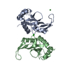
 Components
 Keywords
 LIGASE / RING /
  Function and homology information
 Authors
 Singapore, 1items
  Citation
 Structure visualization
 Downloads & links
 Other downloads
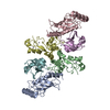


 PDBj
PDBj

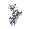
 Assembly
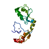


 Mass: 65.409 Da / Num. of mol.: 4 / Source method: obtained synthetically / Formula: Zn
 Mass: 18.015 Da / Num. of mol.: 96 / Source method: isolated from a natural source / Formula: H2O
 Sample preparation
 / Beamline: 24-ID-C / Wavelength: 0.987 Å
 Processing