 Movie
Movie Controller

[English] 日本語
 Yorodumi
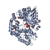
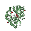
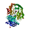
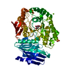
- PDB-5h71: Structure of alginate-binding protein AlgQ2 in complex with an al... -
+ Open data

- Basic information
| Entry | Database: PDB / ID: 5h71 | |||||||||
|---|---|---|---|---|---|---|---|---|---|---|
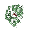
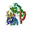
| Title | Structure of alginate-binding protein AlgQ2 in complex with an alginate trisaccharide | |||||||||
 Components Components | AlgQ2 | |||||||||
 Keywords Keywords | SUGAR BINDING PROTEIN / Alginate oligosaccharide / closed conformation / Solute-binding protein /  Sphingomonas Sphingomonas | |||||||||
| Function / homology |  Function and homology information Function and homology information | |||||||||
| Biological species |  Sphingomonas sp. (bacteria) Sphingomonas sp. (bacteria) | |||||||||
| Method | X-RAY DIFFRACTION / SYNCHROTRON / MOLECULAR REPLACEMENT / Resolution: 1.549 Å | |||||||||
 Authors Authors | Uenishi, K. / Kaneko, A. / Maruyama, Y. / Mikami, B. / Murata, K. / Hashimoto, W. | |||||||||
| Funding support |  Japan, 1items Japan, 1items
| |||||||||
 Citation Citation | Journal: J. Biol. Chem. / Year: 2017 Title: A solute-binding protein in the closed conformation induces ATP hydrolysis in a bacterial ATP-binding cassette transporter involved in the import of alginate Authors: Kaneko, A. / Uenishi, K. / Maruyama, Y. / Mizuno, N. / Baba, S. / Kumasaka, T. / Mikami, B. / Murata, K. / Hashimoto, W. | |||||||||
| History |
|
- Structure visualization
| Structure viewer | Molecule: MolmilJmol/JSmol |
|---|
- Downloads & links
-Download
| PDBx/mmCIF format | 5h71.cif.gz | 250.6 KB | Display | PDBx/mmCIF format |
|---|---|---|---|---|
| PDB format | pdb5h71.ent.gz | 196.4 KB | Display | PDB format |
| PDBx/mmJSON format | 5h71.json.gz | Tree view | PDBx/mmJSON format | |
| Others |  Other downloads Other downloads |
-Validation report
| Arichive directory | https://data.pdbj.org/pub/pdb/validation_reports/h7/5h71ftp://data.pdbj.org/pub/pdb/validation_reports/h7/5h71 | HTTPS FTP |
|---|
-Related structure data
| Related structure data | 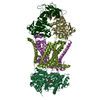 4xigC 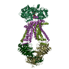 4xtcC  5h6uC  1j1nS C: citing same article ( S: Starting model for refinement |
|---|---|
| Similar structure data |

-Links
 PDBj
PDBj


- Assembly
| Deposited unit | 
| ||||||||
|---|---|---|---|---|---|---|---|---|---|
| 1 | 
| ||||||||
| 2 | 
| ||||||||
| Unit cell |
|
-Components
-Protein / Sugars , 2 types, 4 molecules AB
| #1: Protein | Mass: 57251.715 Da / Num. of mol.: 2 / Fragment: UNP residues 25-516 / Mutation: R253K Source method: isolated from a genetically manipulated source Source: (gene. exp.) Sphingomonas sp. (bacteria) / Gene: algQ2 / Production host: Escherichia coli (E. coli) / References: UniProt: Q9KWT5#2: Polysaccharide | / Mass: 528.372 Da / Num. of mol.: 2Source method: isolated from a genetically manipulated source |
|---|
-Non-polymers , 4 types, 1479 molecules 


| #3: Chemical |  Mass: 40.078 Da / Num. of mol.: 2 Mass: 40.078 Da / Num. of mol.: 2Source method: isolated from a genetically manipulated source Formula: Ca #4: Chemical | Diethylene glycol Mass: 106.120 Da / Num. of mol.: 2 / Source method: obtained synthetically / Formula: C4H10O3 Mass: 106.120 Da / Num. of mol.: 2 / Source method: obtained synthetically / Formula: C4H10O3#5: Chemical | Chloride Mass: 35.453 Da / Num. of mol.: 2 / Source method: obtained synthetically / Formula: Cl Mass: 35.453 Da / Num. of mol.: 2 / Source method: obtained synthetically / Formula: Cl#6: Water | ChemComp-HOH / | WaterMass: 18.015 Da / Num. of mol.: 1473 / Source method: isolated from a natural source / Formula: H2O |
|---|
-Experimental details
-Experiment
| Experiment | Method: X-RAY DIFFRACTION / Number of used crystals: 1 |
|---|
- Sample preparation
| Crystal | Density Matthews: 2.26 Å3/Da / Density % sol: 45.55 % |
|---|---|
| Crystal grow | Temperature: 293.15 K / Method: vapor diffusion, sitting drop / pH: 8.5 Details: 30% PEG4000, 0.2 M lithium sulfate, 0.1 M tris(hydroxymethyl)aminomethan-hydrochloride acid |
-Data collection
| Diffraction | Mean temperature: 100 K |
|---|---|
| Diffraction source | Source: SYNCHROTRON / Site: SPring-8 / Beamline: BL26B2 / Wavelength: 1 Å |
| Detector | Type: RAYONIX MX225HE / Detector: CCD / Date: Oct 9, 2015 |
| Radiation | Protocol: SINGLE WAVELENGTH / Monochromatic (M) / Laue (L): M / Scattering type: x-ray |
| Radiation wavelength | Wavelength: 1 Å / Relative weight: 1 |
| Reflection | Resolution: 1.549→30 Å / Num. obs: 143400 / % possible obs: 96.8 % / Redundancy: 3.8 % / Rmerge(I) obs: 0.084 / Net I/σ(I): 25.9 |
| Reflection shell | Resolution: 1.549→1.58 Å / Redundancy: 2 % / Rmerge(I) obs: 0.42 / Mean I/σ(I) obs: 3.2 / % possible all: 89.1 |
- Processing
| Software |
| |||||||||||||||||||||||||||||||||||||||||||||||||||||||||||||||||||||||||||||||||||||||||||||||||||||||||||||||||||||||||||||||||||||||||||||||||||||||||||||||||||||||||||||||||||||||||||||||||||||||||||||||||||||||||
|---|---|---|---|---|---|---|---|---|---|---|---|---|---|---|---|---|---|---|---|---|---|---|---|---|---|---|---|---|---|---|---|---|---|---|---|---|---|---|---|---|---|---|---|---|---|---|---|---|---|---|---|---|---|---|---|---|---|---|---|---|---|---|---|---|---|---|---|---|---|---|---|---|---|---|---|---|---|---|---|---|---|---|---|---|---|---|---|---|---|---|---|---|---|---|---|---|---|---|---|---|---|---|---|---|---|---|---|---|---|---|---|---|---|---|---|---|---|---|---|---|---|---|---|---|---|---|---|---|---|---|---|---|---|---|---|---|---|---|---|---|---|---|---|---|---|---|---|---|---|---|---|---|---|---|---|---|---|---|---|---|---|---|---|---|---|---|---|---|---|---|---|---|---|---|---|---|---|---|---|---|---|---|---|---|---|---|---|---|---|---|---|---|---|---|---|---|---|---|---|---|---|---|---|---|---|---|---|---|---|---|---|---|---|---|---|---|---|---|
| Refinement | Method to determine structure: MOLECULAR REPLACEMENT Starting model: 1J1N Resolution: 1.549→29.852 Å / SU ML: 0.14 / Cross valid method: FREE R-VALUE / σ(F): 1.34 / Phase error: 19.4
| |||||||||||||||||||||||||||||||||||||||||||||||||||||||||||||||||||||||||||||||||||||||||||||||||||||||||||||||||||||||||||||||||||||||||||||||||||||||||||||||||||||||||||||||||||||||||||||||||||||||||||||||||||||||||
| Solvent computation | Shrinkage radii: 0.9 Å / VDW probe radii: 1.11 Å | |||||||||||||||||||||||||||||||||||||||||||||||||||||||||||||||||||||||||||||||||||||||||||||||||||||||||||||||||||||||||||||||||||||||||||||||||||||||||||||||||||||||||||||||||||||||||||||||||||||||||||||||||||||||||
| Refinement step | Cycle: LAST / Resolution: 1.549→29.852 Å
| |||||||||||||||||||||||||||||||||||||||||||||||||||||||||||||||||||||||||||||||||||||||||||||||||||||||||||||||||||||||||||||||||||||||||||||||||||||||||||||||||||||||||||||||||||||||||||||||||||||||||||||||||||||||||
| Refine LS restraints |
| |||||||||||||||||||||||||||||||||||||||||||||||||||||||||||||||||||||||||||||||||||||||||||||||||||||||||||||||||||||||||||||||||||||||||||||||||||||||||||||||||||||||||||||||||||||||||||||||||||||||||||||||||||||||||
| LS refinement shell |
|
