 Movie
Movie Controller


+ Open data

- Basic information
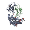
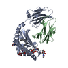
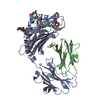
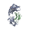
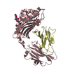
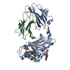
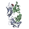
| Entry | Database: PDB / ID: 5h5z | |||||||||
|---|---|---|---|---|---|---|---|---|---|---|
| Title | Crystal structure of bony fish MHC class I, peptide and B2m II | |||||||||
 Components Components |
| |||||||||
 Keywords Keywords | PEPTIDE BINDING PROTEIN/IMMUNE SYSTEM / MHC /  IMMUNOLOGY / IMMUNE SYSTEM-TRANSRERASE COMPLEX / PEPTIDE BINDING PROTEIN-IMMUNE SYSTEM complex IMMUNOLOGY / IMMUNE SYSTEM-TRANSRERASE COMPLEX / PEPTIDE BINDING PROTEIN-IMMUNE SYSTEM complex | |||||||||
| Function / homology |  Function and homology information Function and homology informationNNS virus cap methyltransferase / GDP polyribonucleotidyltransferase / negative stranded viral RNA replication / antigen processing and presentation of peptide antigen via MHC class I / viral transcription / Hydrolases; Acting on acid anhydrides; In phosphorus-containing anhydrides / virion component / MHC class I protein complex / mRNA 5'-cap (guanine-N7-)-methyltransferase activity / host cell cytoplasm ...NNS virus cap methyltransferase / GDP polyribonucleotidyltransferase / negative stranded viral RNA replication / antigen processing and presentation of peptide antigen via MHC class I / viral transcription / Hydrolases; Acting on acid anhydrides; In phosphorus-containing anhydrides / virion component / MHC class I protein complex / mRNA 5'-cap (guanine-N7-)-methyltransferase activity / host cell cytoplasm / immune response / RNA-directed RNA polymerase / RNA-dependent RNA polymerase activity / GTPase activity / extracellular region / ATP binding / membraneSimilarity search - Function | |||||||||
| Biological species |  Ctenopharyngodon idella (grass carp) Ctenopharyngodon idella (grass carp) Spring viraemia of carp virus Spring viraemia of carp virus | |||||||||
| Method | X-RAY DIFFRACTION / MOLECULAR REPLACEMENT / Resolution: 1.74 Å | |||||||||
 Authors Authors | Chen, Z. / Zhang, N. / Qi, J. / Li, X. / Chen, R. / Wang, Z. / Gao, F.G. / Xia, C. | |||||||||
| Funding support |  China, 2items China, 2items
| |||||||||
 Citation Citation | Journal: Iscience / Year: 2020 Title: The Mechanism of beta 2m Molecule-Induced Changes in the Peptide Presentation Profile in a Bony Fish. Authors: Li, Z. / Zhang, N. / Ma, L. / Zhang, L. / Meng, G. / Xia, C. | |||||||||
| History |
|
- Structure visualization
| Structure viewer | Molecule: MolmilJmol/JSmol |
|---|
- Downloads & links
-Download
| PDBx/mmCIF format | 5h5z.cif.gz | 185.8 KB | Display | PDBx/mmCIF format |
|---|---|---|---|---|
| PDB format | pdb5h5z.ent.gz | 145.7 KB | Display | PDB format |
| PDBx/mmJSON format | 5h5z.json.gz | Tree view | PDBx/mmJSON format | |
| Others |  Other downloads Other downloads |
-Validation report
| Arichive directory | https://data.pdbj.org/pub/pdb/validation_reports/h5/5h5zftp://data.pdbj.org/pub/pdb/validation_reports/h5/5h5z | HTTPS FTP |
|---|
-Related structure data
| Related structure data |  6lbeC 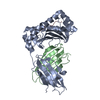 4e0rS S: Starting model for refinement C: citing same article ( |
|---|---|
| Similar structure data |


-Links
 PDBj
PDBj
- Assembly
| Deposited unit | 
| ||||||||
|---|---|---|---|---|---|---|---|---|---|
| 1 |
| ||||||||
| Unit cell |
|
-Components
| #1: Protein | Mass: 31449.723 Da / Num. of mol.: 1 / Fragment: UNP RESIDUES 17-291 / Mutation: E45K Source method: isolated from a genetically manipulated source Source: (gene. exp.) Ctenopharyngodon idella (grass carp) / Gene: UAA106 / Production host:  Escherichia coli (E. coli) / References: UniProt: Q65XY8 Escherichia coli (E. coli) / References: UniProt: Q65XY8 |
|---|---|
| #2: Protein | Beta-2 microglobulin Mass: 11315.831 Da / Num. of mol.: 1 Source method: isolated from a genetically manipulated source Source: (gene. exp.) Ctenopharyngodon idella (grass carp) / Gene: B2m / Production host: Escherichia coli (E. coli) / References: UniProt: Q6L7B0 |
| #3: Protein/peptide | Mass: 1089.393 Da / Num. of mol.: 1 / Source method: obtained synthetically / Source: (synth.) Spring viraemia of carp virus / References: UniProt: Q91DR9*PLUS |
| #4: Water | ChemComp-HOH / Water Mass: 18.015 Da / Num. of mol.: 351 / Source method: isolated from a natural source / Formula: H2O Mass: 18.015 Da / Num. of mol.: 351 / Source method: isolated from a natural source / Formula: H2O |
-Experimental details
-Experiment
| Experiment | Method: X-RAY DIFFRACTION / Number of used crystals: 1 |
|---|
- Sample preparation
| Crystal | Density Matthews: 2.41 Å3/Da / Density % sol: 48.88 % |
|---|---|
| Crystal grow | Temperature: 291 K / Method: vapor diffusion, sitting drop Details: 0.2 M Sodium citrate tribasic dihydrate, 20% w/v Polyethylene glycol 3350 |
-Data collection
| Diffraction | Mean temperature: 100 K |
|---|---|
| Diffraction source | Source: ROTATING ANODE / Type: RIGAKU MICROMAX-007 HF / Wavelength: 0.9792 Å |
| Detector | Type: RIGAKU RAXIS IV++ / Detector: IMAGE PLATE / Date: Jan 1, 2015 |
| Radiation | Protocol: SINGLE WAVELENGTH / Monochromatic (M) / Laue (L): M / Scattering type: x-ray |
| Radiation wavelength | Wavelength: 0.9792 Å / Relative weight: 1 |
| Reflection | Resolution: 1.74→50 Å / Num. obs: 42201 / % possible obs: 98.5 % / Redundancy: 3.6 % / Net I/σ(I): 17.5 |
| Reflection shell | Resolution: 1.74→1.79 Å / Mean I/σ(I) obs: 3 / % possible all: 98.3 |
- Processing
| Software |
| ||||||||||||||||||||||||||||||||||||||||||||||||||||||||||||||||||||||||||||||||||||||||||||||||||||||||||||||||||||||||||||||||||||||||||||||||||||||||||||||||||||||||||||||||||||||
|---|---|---|---|---|---|---|---|---|---|---|---|---|---|---|---|---|---|---|---|---|---|---|---|---|---|---|---|---|---|---|---|---|---|---|---|---|---|---|---|---|---|---|---|---|---|---|---|---|---|---|---|---|---|---|---|---|---|---|---|---|---|---|---|---|---|---|---|---|---|---|---|---|---|---|---|---|---|---|---|---|---|---|---|---|---|---|---|---|---|---|---|---|---|---|---|---|---|---|---|---|---|---|---|---|---|---|---|---|---|---|---|---|---|---|---|---|---|---|---|---|---|---|---|---|---|---|---|---|---|---|---|---|---|---|---|---|---|---|---|---|---|---|---|---|---|---|---|---|---|---|---|---|---|---|---|---|---|---|---|---|---|---|---|---|---|---|---|---|---|---|---|---|---|---|---|---|---|---|---|---|---|---|---|
| Refinement | Method to determine structure: MOLECULAR REPLACEMENT Starting model: 4E0R Resolution: 1.74→50 Å / Cor.coef. Fo:Fc: 0.963 / Cor.coef. Fo:Fc free: 0.949 / SU B: 6.272 / SU ML: 0.088 / Cross valid method: NONE / ESU R: 0.186 / ESU R Free: 0.106
| ||||||||||||||||||||||||||||||||||||||||||||||||||||||||||||||||||||||||||||||||||||||||||||||||||||||||||||||||||||||||||||||||||||||||||||||||||||||||||||||||||||||||||||||||||||||
| Solvent computation | Ion probe radii: 0.8 Å / Shrinkage radii: 0.8 Å / VDW probe radii: 1.2 Å | ||||||||||||||||||||||||||||||||||||||||||||||||||||||||||||||||||||||||||||||||||||||||||||||||||||||||||||||||||||||||||||||||||||||||||||||||||||||||||||||||||||||||||||||||||||||
| Displacement parameters | Biso mean: 32.855 Å2
| ||||||||||||||||||||||||||||||||||||||||||||||||||||||||||||||||||||||||||||||||||||||||||||||||||||||||||||||||||||||||||||||||||||||||||||||||||||||||||||||||||||||||||||||||||||||
| Refinement step | Cycle: 1 / Resolution: 1.74→50 Å
| ||||||||||||||||||||||||||||||||||||||||||||||||||||||||||||||||||||||||||||||||||||||||||||||||||||||||||||||||||||||||||||||||||||||||||||||||||||||||||||||||||||||||||||||||||||||
| Refine LS restraints |
|