 Movie
Movie Controller

[English] 日本語
 Yorodumi
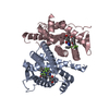
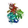
- PDB-5h5j: Complex between ferredoxin and ferredoxin-NADP+ reductase from ma... -
+ Open data

- Basic information
| Entry | Database: PDB / ID: 5h5j | ||||||
|---|---|---|---|---|---|---|---|
| Title | Complex between ferredoxin and ferredoxin-NADP+ reductase from maize root | ||||||
 Components Components |
| ||||||
 Keywords Keywords | OXIDOREDUCTASE/ELECTRON TRANSPORT /  Electron transfer complex / OXIDOREDUCTASE-ELECTRON TRANSPORT complex Electron transfer complex / OXIDOREDUCTASE-ELECTRON TRANSPORT complex | ||||||
| Function / homology |  Function and homology information Function and homology informationetioplast stroma / oxidoreductase activity, acting on NAD(P)H, heme protein as acceptor / ferredoxin-NADP+ reductase / ferredoxin-NADP+ reductase activity / NADPH dehydrogenase activity / chloroplast stroma / NADPH binding / photosynthesis / chloroplast / electron transport chain ...etioplast stroma / oxidoreductase activity, acting on NAD(P)H, heme protein as acceptor / ferredoxin-NADP+ reductase / ferredoxin-NADP+ reductase activity / NADPH dehydrogenase activity / chloroplast stroma / NADPH binding / photosynthesis / chloroplast / electron transport chain / 2 iron, 2 sulfur cluster binding / electron transfer activity / nucleotide binding / metal ion bindingSimilarity search - Function | ||||||
| Biological species |  Zea mays (maize) Zea mays (maize) | ||||||
| Method | X-RAY DIFFRACTION / SYNCHROTRON / MOLECULAR REPLACEMENT / Resolution: 2.5 Å | ||||||
 Authors Authors | Kurisu, G. / Hase, T. | ||||||
| Funding support |  Japan, 1items Japan, 1items
| ||||||
 Citation Citation | Journal: Photosyn. Res. / Year: 2017 Title: Structural basis for the isotype-specific interactions of ferredoxin and ferredoxin: NADP(+) oxidoreductase: an evolutionary switch between photosynthetic and heterotrophic assimilation Authors: Shinohara, F. / Kurisu, G. / Hanke, G. / Bowsher, C. / Hase, T. / Kimata-Ariga, Y. #1: Journal: Photosyn. Res. / Year: 2004 Title: Fd : FNR Electron Transfer Complexes: Evolutionary Refinement of Structural Interactions Authors: Hanke, G.T. / Kurisu, G. / Kusunoki, M. / Hase, T. | ||||||
| History |
|
- Structure visualization
| Structure viewer | Molecule: MolmilJmol/JSmol |
|---|
- Downloads & links
-Download
| PDBx/mmCIF format | 5h5j.cif.gz | 163.4 KB | Display | PDBx/mmCIF format |
|---|---|---|---|---|
| PDB format | pdb5h5j.ent.gz | 125.3 KB | Display | PDB format |
| PDBx/mmJSON format | 5h5j.json.gz | Tree view | PDBx/mmJSON format | |
| Others |  Other downloads Other downloads |
-Validation report
| Arichive directory | https://data.pdbj.org/pub/pdb/validation_reports/h5/5h5jftp://data.pdbj.org/pub/pdb/validation_reports/h5/5h5j | HTTPS FTP |
|---|
-Related structure data
| Related structure data |  5h57C 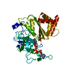 5h59SC C: citing same article ( S: Starting model for refinement |
|---|---|
| Similar structure data |


-Links
 PDBj
PDBj


- Assembly
| Deposited unit | 
| ||||||||
|---|---|---|---|---|---|---|---|---|---|
| 1 | 
| ||||||||
| 2 | 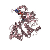
| ||||||||
| Unit cell |
|
-Components
| #1: Protein | Mass: 34632.242 Da / Num. of mol.: 2 / Fragment: UNP residues 19-327 Source method: isolated from a genetically manipulated source Source: (gene. exp.) Zea mays (maize) / Production host:  Escherichia coli (E. coli) Escherichia coli (E. coli)References: UniProt: Q41736, UniProt: B4G043*PLUS, ferredoxin-NADP+ reductase#2: Protein | | / Ferredoxin III / Fd IIIMass: 10362.236 Da / Num. of mol.: 1 Source method: isolated from a genetically manipulated source Source: (gene. exp.) Zea mays (maize) / Gene: FDX3, PFD3 / Production host: Escherichia coli (E. coli) / References: UniProt: P27788#3: Chemical | Flavin adenine dinucleotide  Mass: 785.550 Da / Num. of mol.: 2 Mass: 785.550 Da / Num. of mol.: 2Source method: isolated from a genetically manipulated source Formula: C27H33N9O15P2 / Comment: FAD*YM #4: Chemical | ChemComp-FES / | Iron–sulfur cluster  Mass: 175.820 Da / Num. of mol.: 1 Mass: 175.820 Da / Num. of mol.: 1Source method: isolated from a genetically manipulated source Formula: Fe2S2 #5: Water | ChemComp-HOH / | Water Mass: 18.015 Da / Num. of mol.: 334 / Source method: isolated from a natural source / Formula: H2O Mass: 18.015 Da / Num. of mol.: 334 / Source method: isolated from a natural source / Formula: H2O |
|---|
-Experimental details
-Experiment
| Experiment | Method: X-RAY DIFFRACTION / Number of used crystals: 1 |
|---|
- Sample preparation
| Crystal | Density Matthews: 2.25 Å3/Da / Density % sol: 45.41 % |
|---|---|
| Crystal grow | Temperature: 277 K / Method: vapor diffusion, hanging drop / pH: 6.5 / Details: 20 % PEG6000, 100 mM Sodium cacodylate |
-Data collection
| Diffraction | Mean temperature: 100 K |
|---|---|
| Diffraction source | Source: SYNCHROTRON / Site: SPring-8 / Beamline: BL41XU / Wavelength: 1 Å |
| Detector | Type: ADSC QUANTUM 4 / Detector: CCD / Date: Dec 18, 2000 |
| Radiation | Protocol: SINGLE WAVELENGTH / Monochromatic (M) / Laue (L): M / Scattering type: x-ray |
| Radiation wavelength | Wavelength: 1 Å / Relative weight: 1 |
| Reflection | Resolution: 2.49→50 Å / Num. obs: 91157 / % possible obs: 99.4 % / Redundancy: 3.74 % / Biso Wilson estimate: 19.9 Å2 / Net I/σ(I): 25.7 |
- Processing
| Software |
| ||||||||||||||||||||||||||||||||||||||||||||||||||||||||||||||||||||||||||||||||
|---|---|---|---|---|---|---|---|---|---|---|---|---|---|---|---|---|---|---|---|---|---|---|---|---|---|---|---|---|---|---|---|---|---|---|---|---|---|---|---|---|---|---|---|---|---|---|---|---|---|---|---|---|---|---|---|---|---|---|---|---|---|---|---|---|---|---|---|---|---|---|---|---|---|---|---|---|---|---|---|---|---|
| Refinement | Method to determine structure: MOLECULAR REPLACEMENT Starting model: 5H59 Resolution: 2.5→19.89 Å / Rfactor Rfree error: 0.008 / Data cutoff high absF: 4560843.87 / Data cutoff low absF: 0 / Cross valid method: THROUGHOUT / σ(F): 2 / Details: RESOLUTION-DEPENDENT WEIGHTING SCHEME USE
| ||||||||||||||||||||||||||||||||||||||||||||||||||||||||||||||||||||||||||||||||
| Solvent computation | Bsol: 35.577 Å2 / ksol: 0.370307 e/Å3 | ||||||||||||||||||||||||||||||||||||||||||||||||||||||||||||||||||||||||||||||||
| Displacement parameters | Biso mean: 21.2 Å2
| ||||||||||||||||||||||||||||||||||||||||||||||||||||||||||||||||||||||||||||||||
| Refine analyze |
| ||||||||||||||||||||||||||||||||||||||||||||||||||||||||||||||||||||||||||||||||
| Refinement step | Cycle: 1 / Resolution: 2.5→19.89 Å
| ||||||||||||||||||||||||||||||||||||||||||||||||||||||||||||||||||||||||||||||||
| Refine LS restraints |
| ||||||||||||||||||||||||||||||||||||||||||||||||||||||||||||||||||||||||||||||||
| LS refinement shell | Resolution: 2.5→2.66 Å / Rfactor Rfree error: 0.021 / Total num. of bins used: 6
| ||||||||||||||||||||||||||||||||||||||||||||||||||||||||||||||||||||||||||||||||
| Xplor file |
|
