 Movie
Movie Controller

[English] 日本語
 Yorodumi
- PDB-5h53: The structure of rabbit skeletal muscle actomyosin rigor complex ... -
+ Open data

- Basic information
| Entry | Database: PDB / ID: 5h53 | ||||||
|---|---|---|---|---|---|---|---|
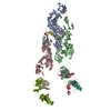
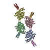
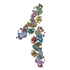
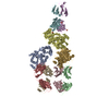
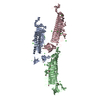
| Title | The structure of rabbit skeletal muscle actomyosin rigor complex at 5.2 angstrom. | ||||||
 Components Components |
| ||||||
 Keywords Keywords |  MOTOR PROTEIN / Actin / Myosin / Muscle / rigor complex MOTOR PROTEIN / Actin / Myosin / Muscle / rigor complex | ||||||
| Function / homology |  Function and homology informationmyosin complex / structural constituent of muscle / cytoskeletal motor activator activity / tropomyosin binding / myofibril / myosin heavy chain binding / mesenchyme migration / troponin I binding / actin filament bundle / filamentous actin ...myosin complex / structural constituent of muscle / cytoskeletal motor activator activity / tropomyosin binding / myofibril / myosin heavy chain binding / mesenchyme migration / troponin I binding / actin filament bundle / filamentous actin / actin filament bundle assembly / skeletal muscle thin filament assembly / cytoskeletal motor activity / striated muscle thin filament / skeletal muscle myofibril / actin monomer binding / skeletal muscle fiber development / stress fiber / titin binding / actin filament polymerization / cellular response to starvation / filopodium / actin filament / Hydrolases; Acting on acid anhydrides; Acting on acid anhydrides to facilitate cellular and subcellular movement / calcium-dependent protein binding / actin filament binding / lamellipodium / cell body / hydrolase activity / protein domain specific binding / calcium ion binding / positive regulation of gene expression / magnesium ion binding / ATP binding / identical protein binding / cytoplasm Function and homology informationmyosin complex / structural constituent of muscle / cytoskeletal motor activator activity / tropomyosin binding / myofibril / myosin heavy chain binding / mesenchyme migration / troponin I binding / actin filament bundle / filamentous actin ...myosin complex / structural constituent of muscle / cytoskeletal motor activator activity / tropomyosin binding / myofibril / myosin heavy chain binding / mesenchyme migration / troponin I binding / actin filament bundle / filamentous actin / actin filament bundle assembly / skeletal muscle thin filament assembly / cytoskeletal motor activity / striated muscle thin filament / skeletal muscle myofibril / actin monomer binding / skeletal muscle fiber development / stress fiber / titin binding / actin filament polymerization / cellular response to starvation / filopodium / actin filament / Hydrolases; Acting on acid anhydrides; Acting on acid anhydrides to facilitate cellular and subcellular movement / calcium-dependent protein binding / actin filament binding / lamellipodium / cell body / hydrolase activity / protein domain specific binding / calcium ion binding / positive regulation of gene expression / magnesium ion binding / ATP binding / identical protein binding / cytoplasmSimilarity search - Function | ||||||
| Biological species |  Oryctolagus cuniculus (rabbit) Oryctolagus cuniculus (rabbit) | ||||||
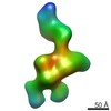
| Method | ELECTRON MICROSCOPY / helical reconstruction / cryo EM / Resolution: 5.2 Å | ||||||
 Authors Authors | Fujii, T. / Namba, K. | ||||||
 Citation Citation | Journal: Nat Commun / Year: 2017 Title: Structure of actomyosin rigour complex at 5.2 Å resolution and insights into the ATPase cycle mechanism. Authors: Takashi Fujii / Keiichi Namba /  Abstract: Muscle contraction is driven by cyclic association and dissociation of myosin head of the thick filament with thin actin filament coupled with ATP binding and hydrolysis by myosin. However, because ...Muscle contraction is driven by cyclic association and dissociation of myosin head of the thick filament with thin actin filament coupled with ATP binding and hydrolysis by myosin. However, because of the absence of actomyosin rigour structure at high resolution, it still remains unclear how the strong binding of myosin to actin filament triggers the release of hydrolysis products and how ATP binding causes their dissociation. Here we report the structure of mammalian skeletal muscle actomyosin rigour complex at 5.2 Å resolution by electron cryomicroscopy. Comparison with the structures of myosin in various states shows a distinctly large conformational change, providing insights into the ATPase-coupled reaction cycle of actomyosin. Based on our observations, we hypothesize that asymmetric binding along the actin filament could function as a Brownian ratchet by favouring directionally biased thermal motions of myosin and actin. | ||||||
| History |
|
- Structure visualization
| Movie |
Movie viewer |
|---|---|
| Structure viewer | Molecule: MolmilJmol/JSmol |
- Downloads & links
-Download
| PDBx/mmCIF format | 5h53.cif.gz | 328.6 KB | Display | PDBx/mmCIF format |
|---|---|---|---|---|
| PDB format | pdb5h53.ent.gz | 265.6 KB | Display | PDB format |
| PDBx/mmJSON format | 5h53.json.gz | Tree view | PDBx/mmJSON format | |
| Others |  Other downloads Other downloads |
-Validation report
| Arichive directory | https://data.pdbj.org/pub/pdb/validation_reports/h5/5h53ftp://data.pdbj.org/pub/pdb/validation_reports/h5/5h53 | HTTPS FTP |
|---|
-Related structure data
| Related structure data | 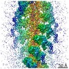 6664MC M: map data used to model this data C: citing same article ( |
|---|---|
| Similar structure data |
-Links
 PDBj
PDBj


- Assembly
| Deposited unit | 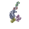
|
|---|---|
| 1 |
|
-Components
| #1: Protein | Mass: 96770.133 Da / Num. of mol.: 1 / Fragment: UNP residues 1-845 / Source method: isolated from a natural source / Source: (natural) Oryctolagus cuniculus (rabbit) / References: UniProt: Q9GJP9 | ||
|---|---|---|---|
| #2: Protein | Mass: 16507.588 Da / Num. of mol.: 1 / Fragment: UNP residues 25-170 / Source method: isolated from a natural source / Source: (natural) Oryctolagus cuniculus (rabbit) / References: UniProt: P24732 | ||
| #3: Protein | Mass: 17187.293 Da / Num. of mol.: 1 / Fragment: UNP residues 41-192 / Source method: isolated from a natural source / Source: (natural) Oryctolagus cuniculus (rabbit) / References: UniProt: P02602 | ||
| #4: Protein | / Alpha-actin-1 Mass: 41875.633 Da / Num. of mol.: 2 / Fragment: UNP residues 3-377 / Source method: isolated from a natural source / Source: (natural) Oryctolagus cuniculus (rabbit) / References: UniProt: P68135#5: Chemical | Adenosine diphosphate  Mass: 427.201 Da / Num. of mol.: 2 / Source method: obtained synthetically / Formula: C10H15N5O10P2 / Comment: ADP, energy-carrying molecule*YM Mass: 427.201 Da / Num. of mol.: 2 / Source method: obtained synthetically / Formula: C10H15N5O10P2 / Comment: ADP, energy-carrying molecule*YM |
-Experimental details
-Experiment
| Experiment | Method: ELECTRON MICROSCOPY |
|---|---|
| EM experiment | Aggregation state: FILAMENT / 3D reconstruction method: helical reconstruction |
- Sample preparation
| Component | Name: Actomyosin rigor complex / Type: COMPLEX / Entity ID: #1-#4 / Source: NATURAL |
|---|---|
| Source (natural) | Organism: Oryctolagus cuniculus (rabbit) |
| Buffer solution | pH: 7.5 |
| Specimen | Embedding applied: NO / Shadowing applied: NO / Staining applied: NO / Vitrification applied: YES |
| Vitrification | Cryogen name: ETHANE |
- Electron microscopy imaging
| Microscopy | Model: JEOL 3200FSC |
|---|---|
| Electron gun | Electron source: FIELD EMISSION GUN / Accelerating voltage: 200 kV / Illumination mode: FLOOD BEAM |
| Electron lens | Mode: BRIGHT FIELDBright-field microscopy |
| Image recording | Electron dose: 20 e/Å2 / Film or detector model: TVIPS TEMCAM-F415 (4k x 4k) |
- Processing
| CTF correction | Type: PHASE FLIPPING AND AMPLITUDE CORRECTION |
|---|---|
| Helical symmerty | Angular rotation/subunit: 166.7 ° / Axial rise/subunit: 27.6 Å / Axial symmetry: C1 |
| 3D reconstruction | Resolution: 5.2 Å / Resolution method: FSC 0.143 CUT-OFF / Num. of particles: 31535 / Symmetry type: HELICAL |
