 Movie
Movie Controller

[English] 日本語
 Yorodumi
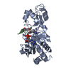
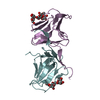
- PDB-5h3j: Crystal structure of Grasp domain of Grasp55 complexed with the G... -
+ Open data

- Basic information
| Entry | Database: PDB / ID: 5h3j | ||||||
|---|---|---|---|---|---|---|---|
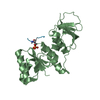
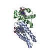
| Title | Crystal structure of Grasp domain of Grasp55 complexed with the Golgin45 C-terminus | ||||||
 Components Components |
| ||||||
 Keywords Keywords |  PROTEIN TRANSPORT / PDZ DOMAIN / GOLGI peripheral membrane PROTEIN / GOLGIN / TETHERING PROTEIN PROTEIN TRANSPORT / PDZ DOMAIN / GOLGI peripheral membrane PROTEIN / GOLGIN / TETHERING PROTEIN | ||||||
| Function / homology |  Function and homology information Function and homology informationGolgi Cisternae Pericentriolar Stack Reorganization / establishment of protein localization to plasma membrane / organelle assembly / organelle organization / Golgi to plasma membrane protein transport / Golgi medial cisterna / Golgi organization / response to endoplasmic reticulum stress / spermatogenesis / cell differentiation ...Golgi Cisternae Pericentriolar Stack Reorganization / establishment of protein localization to plasma membrane / organelle assembly / organelle organization / Golgi to plasma membrane protein transport / Golgi medial cisterna / Golgi organization / response to endoplasmic reticulum stress / spermatogenesis / cell differentiation / Golgi membrane / ubiquitin protein ligase binding / endoplasmic reticulum membrane / Golgi apparatus / enzyme binding / nucleoplasm / nucleusSimilarity search - Function | ||||||
| Biological species |  Mus musculus (house mouse) Mus musculus (house mouse) | ||||||
| Method | X-RAY DIFFRACTION / SYNCHROTRON / MOLECULAR REPLACEMENT / Resolution: 1.33 Å | ||||||
 Authors Authors | Shi, N. / Zhao, J. / Li, B. | ||||||
| Funding support |  China, 1items China, 1items
| ||||||
 Citation Citation | Journal: J. Biol. Chem. / Year: 2017 Title: Structural Basis for the Interaction between Golgi Reassembly-stacking Protein GRASP55 and Golgin45 Authors: Zhao, J. / Li, B. / Huang, X. / Morelli, X. / Shi, N. | ||||||
| History |
|
- Structure visualization
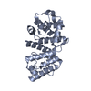
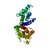
| Structure viewer | Molecule: MolmilJmol/JSmol |
|---|
- Downloads & links
-Download
| PDBx/mmCIF format | 5h3j.cif.gz | 145.5 KB | Display | PDBx/mmCIF format |
|---|---|---|---|---|
| PDB format | pdb5h3j.ent.gz | 114.7 KB | Display | PDB format |
| PDBx/mmJSON format | 5h3j.json.gz | Tree view | PDBx/mmJSON format | |
| Others |  Other downloads Other downloads |
-Validation report
| Arichive directory | https://data.pdbj.org/pub/pdb/validation_reports/h3/5h3jftp://data.pdbj.org/pub/pdb/validation_reports/h3/5h3j | HTTPS FTP |
|---|
-Related structure data
| Related structure data |  3rleS S: Starting model for refinement |
|---|---|
| Similar structure data |


-Links
 PDBj
PDBj- Assembly
| Deposited unit | 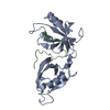
| |||||||||
|---|---|---|---|---|---|---|---|---|---|---|
| 1 |
| |||||||||
| Unit cell |
| |||||||||
| Components on special symmetry positions |
|
-Components
| #1: Protein | Mass: 23029.691 Da / Num. of mol.: 1 / Fragment: Grasp domain, UNP residues 2-208 Source method: isolated from a genetically manipulated source Source: (gene. exp.) Mus musculus (house mouse) / Gene: Gorasp2 / Production host:  Escherichia coli (E. coli) / References: UniProt: Q99JX3 Escherichia coli (E. coli) / References: UniProt: Q99JX3 |
|---|---|
| #2: Protein/peptide | BLZF1 / Basic leucine zipper nuclear factor 1 Mass: 3273.655 Da / Num. of mol.: 1 / Fragment: UNP residues 379-403 Source method: isolated from a genetically manipulated source Source: (gene. exp.) Mus musculus (house mouse) / Gene: Blzf1 / Production host: Escherichia coli (E. coli) / References: UniProt: Q8R2X8 |
| #3: Chemical | ChemComp-ZN /   Mass: 65.409 Da / Num. of mol.: 1 / Source method: obtained synthetically / Formula: Zn Mass: 65.409 Da / Num. of mol.: 1 / Source method: obtained synthetically / Formula: Zn |
| #4: Water | ChemComp-HOH / Water Mass: 18.015 Da / Num. of mol.: 386 / Source method: isolated from a natural source / Formula: H2O Mass: 18.015 Da / Num. of mol.: 386 / Source method: isolated from a natural source / Formula: H2O |
-Experimental details
-Experiment
| Experiment | Method: X-RAY DIFFRACTION / Number of used crystals: 1 |
|---|
- Sample preparation
| Crystal | Density Matthews: 1.97 Å3/Da / Density % sol: 42.18 % |
|---|---|
| Crystal grow | Temperature: 291 K / Method: vapor diffusion, sitting drop / pH: 8.5 / Details: PEG4000, potassium sodium tartrate, Tris |
-Data collection
| Diffraction | Mean temperature: 100 K |
|---|---|
| Diffraction source | Source: SYNCHROTRON / Site: SSRF / Beamline: BL17U / Wavelength: 0.97368 Å |
| Detector | Type: ADSC QUANTUM 315 / Detector: CCD / Date: Jun 9, 2016 |
| Radiation | Protocol: SINGLE WAVELENGTH / Monochromatic (M) / Laue (L): M / Scattering type: x-ray |
| Radiation wavelength | Wavelength: 0.97368 Å / Relative weight: 1 |
| Reflection | Resolution: 1.25→33.65 Å / Num. obs: 122914 / % possible obs: 99.81 % / Redundancy: 7.2 % / Net I/σ(I): 8.38 |
| Reflection shell | Resolution: 1.25→1.295 Å |
- Processing
| Software |
| |||||||||||||||||||||||||||||||||||||||||||||||||||||||||||||||||||||||||||||||||||||||||||||||||||||||||||||||||||||||||||||||||||||||||||||||||||||||||||||||||||||||||||||||||||||||||||||||||||||||||||
|---|---|---|---|---|---|---|---|---|---|---|---|---|---|---|---|---|---|---|---|---|---|---|---|---|---|---|---|---|---|---|---|---|---|---|---|---|---|---|---|---|---|---|---|---|---|---|---|---|---|---|---|---|---|---|---|---|---|---|---|---|---|---|---|---|---|---|---|---|---|---|---|---|---|---|---|---|---|---|---|---|---|---|---|---|---|---|---|---|---|---|---|---|---|---|---|---|---|---|---|---|---|---|---|---|---|---|---|---|---|---|---|---|---|---|---|---|---|---|---|---|---|---|---|---|---|---|---|---|---|---|---|---|---|---|---|---|---|---|---|---|---|---|---|---|---|---|---|---|---|---|---|---|---|---|---|---|---|---|---|---|---|---|---|---|---|---|---|---|---|---|---|---|---|---|---|---|---|---|---|---|---|---|---|---|---|---|---|---|---|---|---|---|---|---|---|---|---|---|---|---|---|---|---|---|
| Refinement | Method to determine structure: MOLECULAR REPLACEMENT Starting model: 3RLE Resolution: 1.33→31.954 Å / SU ML: 0.1 / Cross valid method: FREE R-VALUE / σ(F): 1.34 / Phase error: 16.4 / Stereochemistry target values: ML
| |||||||||||||||||||||||||||||||||||||||||||||||||||||||||||||||||||||||||||||||||||||||||||||||||||||||||||||||||||||||||||||||||||||||||||||||||||||||||||||||||||||||||||||||||||||||||||||||||||||||||||
| Solvent computation | Shrinkage radii: 0.9 Å / VDW probe radii: 1.11 Å / Solvent model: FLAT BULK SOLVENT MODEL | |||||||||||||||||||||||||||||||||||||||||||||||||||||||||||||||||||||||||||||||||||||||||||||||||||||||||||||||||||||||||||||||||||||||||||||||||||||||||||||||||||||||||||||||||||||||||||||||||||||||||||
| Refinement step | Cycle: LAST / Resolution: 1.33→31.954 Å
| |||||||||||||||||||||||||||||||||||||||||||||||||||||||||||||||||||||||||||||||||||||||||||||||||||||||||||||||||||||||||||||||||||||||||||||||||||||||||||||||||||||||||||||||||||||||||||||||||||||||||||
| Refine LS restraints |
| |||||||||||||||||||||||||||||||||||||||||||||||||||||||||||||||||||||||||||||||||||||||||||||||||||||||||||||||||||||||||||||||||||||||||||||||||||||||||||||||||||||||||||||||||||||||||||||||||||||||||||
| LS refinement shell |
|
