 Movie
Movie Controller

[English] 日本語
 Yorodumi
- PDB-5h28: Crystal structure of Osh1 ANK domain from Saccharomyces cerevisia -
+ Open data

- Basic information
| Entry | Database: PDB / ID: 5h28 | ||||||
|---|---|---|---|---|---|---|---|
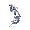
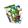
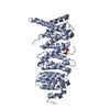
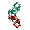
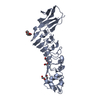
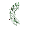
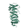
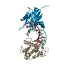
| Title | Crystal structure of Osh1 ANK domain from Saccharomyces cerevisia | ||||||
 Components Components | Oxysterol-binding protein homolog 1 | ||||||
 Keywords Keywords | LIPID BINDING PROTEIN /  oxysterol binding / lipid transfer / ANK Nvj1 oxysterol binding / lipid transfer / ANK Nvj1 | ||||||
| Function / homology |  Function and homology information Function and homology informationSynthesis of bile acids and bile salts / nucleus-vacuole junction / sterol transfer activity / Golgi trans cisterna / : / sterol transport / sterol binding / maintenance of cell polarity / piecemeal microautophagy of the nucleus / nuclear outer membrane ...Synthesis of bile acids and bile salts / nucleus-vacuole junction / sterol transfer activity / Golgi trans cisterna / : / sterol transport / sterol binding / maintenance of cell polarity / piecemeal microautophagy of the nucleus / nuclear outer membrane / vacuolar membrane / exocytosis / endocytosis / nuclear envelope / early endosome / Golgi membrane / intracellular membrane-bounded organelle / lipid binding / endoplasmic reticulum membrane / endoplasmic reticulum / membrane / cytosolSimilarity search - Function | ||||||
| Biological species |  Saccharomyces cerevisiae (brewer's yeast) Saccharomyces cerevisiae (brewer's yeast) | ||||||
| Method | X-RAY DIFFRACTION / SYNCHROTRON / MOLECULAR REPLACEMENT / Resolution: 1.5 Å | ||||||
 Authors Authors | Im, Y.J. / Manik, M.K. / Yang, H.S. / Tong, J.S. | ||||||
| Funding support |  Korea, Republic Of, 1items Korea, Republic Of, 1items
| ||||||
 Citation Citation | Journal: Structure / Year: 2017 Title: Structure of Yeast OSBP-Related Protein Osh1 Reveals Key Determinants for Lipid Transport and Protein Targeting at the Nucleus-Vacuole Junction Authors: Manik, M.K. / Yang, H. / Tong, J. / Im, Y.J. | ||||||
| History |
|
- Structure visualization
| Structure viewer | Molecule: MolmilJmol/JSmol |
|---|
- Downloads & links
-Download
| PDBx/mmCIF format | 5h28.cif.gz | 72.6 KB | Display | PDBx/mmCIF format |
|---|---|---|---|---|
| PDB format | pdb5h28.ent.gz | 51.6 KB | Display | PDB format |
| PDBx/mmJSON format | 5h28.json.gz | Tree view | PDBx/mmJSON format | |
| Others |  Other downloads Other downloads |
-Validation report
| Arichive directory | https://data.pdbj.org/pub/pdb/validation_reports/h2/5h28ftp://data.pdbj.org/pub/pdb/validation_reports/h2/5h28 | HTTPS FTP |
|---|
-Related structure data


-Links
 PDBj
PDBj


- Assembly
| Deposited unit | 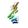
| ||||||||
|---|---|---|---|---|---|---|---|---|---|
| 1 |
| ||||||||
| Unit cell |
|
-Components
| #1: Protein | Mass: 30046.475 Da / Num. of mol.: 1 / Fragment: UNP residues 12-274 Source method: isolated from a genetically manipulated source Source: (gene. exp.) Saccharomyces cerevisiae (strain ATCC 204508 / S288c) (yeast)Strain: ATCC 204508 / S288c / Gene: SWH1, OSH1, YAR042W, YAR044W / Plasmid: modified pHIS-2 / Production host:  Escherichia coli BL21(DE3) (bacteria) / Strain (production host): BL21(DE3) / References: UniProt: P35845 Escherichia coli BL21(DE3) (bacteria) / Strain (production host): BL21(DE3) / References: UniProt: P35845 |
|---|---|
| #2: Water | ChemComp-HOH / Water Mass: 18.015 Da / Num. of mol.: 315 / Source method: isolated from a natural source / Formula: H2O Mass: 18.015 Da / Num. of mol.: 315 / Source method: isolated from a natural source / Formula: H2O |
-Experimental details
-Experiment
| Experiment | Method: X-RAY DIFFRACTION / Number of used crystals: 1 |
|---|
- Sample preparation
| Crystal | Density Matthews: 1.73 Å3/Da / Density % sol: 28.97 % |
|---|---|
| Crystal grow | Temperature: 295 K / Method: vapor diffusion, hanging drop / pH: 7 / Details: 0.1M HEPES-NaOH pH 7.0, 20% PEG 1500 |
-Data collection
| Diffraction | Mean temperature: 100 K |
|---|---|
| Diffraction source | Source: SYNCHROTRON / Site: PAL/PLS / Beamline: 7A (6B, 6C1) / Wavelength: 0.9795 Å |
| Detector | Type: ADSC QUANTUM 270 / Detector: CCD / Date: Jul 5, 2014 / Details: focusing mirror |
| Radiation | Monochromator: Si crystal / Protocol: SINGLE WAVELENGTH / Monochromatic (M) / Laue (L): M / Scattering type: x-ray |
| Radiation wavelength | Wavelength: 0.9795 Å / Relative weight: 1 |
| Reflection | Resolution: 1.5→50 Å / Num. obs: 80061 / % possible obs: 99.6 % / Redundancy: 4.1 % / Biso Wilson estimate: 13.9 Å2 / Rmerge(I) obs: 0.062 / Net I/σ(I): 31.6 |
| Reflection shell | Resolution: 1.5→1.53 Å / Redundancy: 4.1 % / Rmerge(I) obs: 0.371 / Mean I/σ(I) obs: 4.8 / % possible all: 99 |
- Processing
| Software |
| ||||||||||||||||||||||||||||||||||||||||||||||||||||||||||||
|---|---|---|---|---|---|---|---|---|---|---|---|---|---|---|---|---|---|---|---|---|---|---|---|---|---|---|---|---|---|---|---|---|---|---|---|---|---|---|---|---|---|---|---|---|---|---|---|---|---|---|---|---|---|---|---|---|---|---|---|---|---|
| Refinement | Method to determine structure: MOLECULAR REPLACEMENT / Resolution: 1.5→24.55 Å / Rfactor Rfree error: 0.005 / Data cutoff high absF: 693450.38 / Data cutoff low absF: 0 / Cross valid method: THROUGHOUT / σ(F): 0
| ||||||||||||||||||||||||||||||||||||||||||||||||||||||||||||
| Solvent computation | Bsol: 52.4845 Å2 / ksol: 0.37765 e/Å3 | ||||||||||||||||||||||||||||||||||||||||||||||||||||||||||||
| Displacement parameters | Biso mean: 14.3 Å2
| ||||||||||||||||||||||||||||||||||||||||||||||||||||||||||||
| Refine analyze |
| ||||||||||||||||||||||||||||||||||||||||||||||||||||||||||||
| Refinement step | Cycle: 1 / Resolution: 1.5→24.55 Å /
| ||||||||||||||||||||||||||||||||||||||||||||||||||||||||||||
| Refine LS restraints |
| ||||||||||||||||||||||||||||||||||||||||||||||||||||||||||||
| LS refinement shell | Resolution: 1.5→1.59 Å / Rfactor Rfree error: 0.015 / Total num. of bins used: 6
| ||||||||||||||||||||||||||||||||||||||||||||||||||||||||||||
| Xplor file |
|
