+ Open data
- Basic information
| Entry | Database: PDB / ID: 5h1r | ||||||
|---|---|---|---|---|---|---|---|
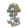
| Title | C. elegans INX-6 gap junction channel | ||||||
 Components Components | Innexin-6 | ||||||
 Keywords Keywords |  TRANSPORT PROTEIN / TRANSPORT PROTEIN /  innexin / gap junction channel / innexin / gap junction channel /  wild type wild type | ||||||
| Function / homology | gap junction hemi-channel activity /  Innexin / Innexin /  Innexin / Pannexin family profile. / Innexin / Pannexin family profile. /  gap junction / gap junction channel activity / monoatomic ion transmembrane transport / gap junction / gap junction channel activity / monoatomic ion transmembrane transport /  plasma membrane / plasma membrane /  Innexin-6 Innexin-6 Function and homology information Function and homology information | ||||||
| Biological species |   Caenorhabditis elegans (invertebrata) Caenorhabditis elegans (invertebrata) | ||||||
| Method |  ELECTRON MICROSCOPY / ELECTRON MICROSCOPY /  single particle reconstruction / single particle reconstruction /  cryo EM / Resolution: 3.6 Å cryo EM / Resolution: 3.6 Å | ||||||
 Authors Authors | Oshima, A. / Tani, K. / Fujiyoshi, Y. | ||||||
 Citation Citation |  Journal: Nat Commun / Year: 2016 Journal: Nat Commun / Year: 2016Title: Atomic structure of the innexin-6 gap junction channel determined by cryo-EM. Authors: Atsunori Oshima / Kazutoshi Tani / Yoshinori Fujiyoshi /  Abstract: Innexins, a large protein family comprising invertebrate gap junction channels, play an essential role in nervous system development and electrical synapse formation. Here we report the cryo-electron ...Innexins, a large protein family comprising invertebrate gap junction channels, play an essential role in nervous system development and electrical synapse formation. Here we report the cryo-electron microscopy structures of Caenorhabditis elegans innexin-6 (INX-6) gap junction channels at atomic resolution. We find that the arrangements of the transmembrane helices and extracellular loops of the INX-6 monomeric structure are highly similar to those of connexin-26 (Cx26), despite the lack of significant sequence similarity. The INX-6 gap junction channel comprises hexadecameric subunits but reveals the N-terminal pore funnel, consistent with Cx26. The helix-rich cytoplasmic loop and C-terminus are intercalated one-by-one through an octameric hemichannel, forming a dome-like entrance that interacts with N-terminal loops in the pore. These observations suggest that the INX-6 cytoplasmic domains are cooperatively associated with the N-terminal funnel conformation, and an essential linkage of the N-terminal with channel activity is presumably preserved across gap junction families. | ||||||
| History |
|
- Structure visualization
| Movie |
 Movie viewer Movie viewer |
|---|---|
| Structure viewer | Molecule:  Molmil Molmil Jmol/JSmol Jmol/JSmol |
- Downloads & links
- Download
| PDBx/mmCIF format |  5h1r.cif.gz 5h1r.cif.gz | 1.1 MB | Display |  PDBx/mmCIF format PDBx/mmCIF format |
|---|---|---|---|---|
| PDB format |  pdb5h1r.ent.gz pdb5h1r.ent.gz | 954.3 KB | Display |  PDB format PDB format |
| PDBx/mmJSON format |  5h1r.json.gz 5h1r.json.gz | Tree view |  PDBx/mmJSON format PDBx/mmJSON format | |
| Others |  Other downloads Other downloads |
-Validation report
| Arichive directory |  https://data.pdbj.org/pub/pdb/validation_reports/h1/5h1r https://data.pdbj.org/pub/pdb/validation_reports/h1/5h1r ftp://data.pdbj.org/pub/pdb/validation_reports/h1/5h1r ftp://data.pdbj.org/pub/pdb/validation_reports/h1/5h1r | HTTPS FTP |
|---|
-Related structure data
| Related structure data |  9571MC  9570C  5h1qC M: map data used to model this data C: citing same article ( |
|---|---|
| Similar structure data |
- Links
Links
- Assembly
| Deposited unit | 
|
|---|---|
| 1 |
|
- Components
| #1: Protein |  / Protein opu-6 / Protein opu-6Mass: 45173.766 Da / Num. of mol.: 16 Source method: isolated from a genetically manipulated source Source: (gene. exp.)   Caenorhabditis elegans (invertebrata) / Gene: inx-6, opu-6, C36H8.2 / Plasmid: pFastBac1 / Cell line (production host): sf9 / Production host: Caenorhabditis elegans (invertebrata) / Gene: inx-6, opu-6, C36H8.2 / Plasmid: pFastBac1 / Cell line (production host): sf9 / Production host:   Spodoptera frugiperda (fall armyworm) / References: UniProt: Q9U3N4 Spodoptera frugiperda (fall armyworm) / References: UniProt: Q9U3N4 |
|---|
-Experimental details
-Experiment
| Experiment | Method:  ELECTRON MICROSCOPY ELECTRON MICROSCOPY |
|---|---|
| EM experiment | Aggregation state: PARTICLE / 3D reconstruction method:  single particle reconstruction single particle reconstruction |
- Sample preparation
| Component | Name: INX-6 / Type: COMPLEX / Entity ID: all / Source: RECOMBINANT |
|---|---|
| Source (natural) | Organism:   Caenorhabditis elegans (invertebrata) Caenorhabditis elegans (invertebrata) |
| Source (recombinant) | Organism:   Spodoptera frugiperda (fall armyworm) / Plasmid Spodoptera frugiperda (fall armyworm) / Plasmid : pFastBac1 : pFastBac1 |
| Buffer solution | pH: 7.5 |
| Specimen | Embedding applied: NO / Shadowing applied: NO / Staining applied : NO / Vitrification applied : NO / Vitrification applied : YES : YES |
Vitrification | Cryogen name: ETHANE |
- Electron microscopy imaging
| Microscopy | Model: JEOL 3000SFF |
|---|---|
| Electron gun | Electron source : :  FIELD EMISSION GUN / Accelerating voltage: 300 kV / Illumination mode: FLOOD BEAM FIELD EMISSION GUN / Accelerating voltage: 300 kV / Illumination mode: FLOOD BEAM |
| Electron lens | Mode: BRIGHT FIELD Bright-field microscopy Bright-field microscopy |
| Image recording | Electron dose: 7 e/Å2 / Film or detector model: GATAN K2 SUMMIT (4k x 4k) |
- Processing
| Software | Name: PHENIX / Version: 1.10.1-2155_1496: / Classification: refinement | ||||||||||||||||||||||||
|---|---|---|---|---|---|---|---|---|---|---|---|---|---|---|---|---|---|---|---|---|---|---|---|---|---|
| EM software | Name: RELION / Version: 1.4 / Category: 3D reconstruction | ||||||||||||||||||||||||
CTF correction | Type: PHASE FLIPPING ONLY | ||||||||||||||||||||||||
| Symmetry | Point symmetry : D8 (2x8 fold dihedral : D8 (2x8 fold dihedral ) ) | ||||||||||||||||||||||||
3D reconstruction | Resolution: 3.6 Å / Resolution method: FSC 0.143 CUT-OFF / Num. of particles: 35608 / Symmetry type: POINT | ||||||||||||||||||||||||
| Refine LS restraints |
|
 Movie
Movie Controller


 PDBj
PDBj