 Movie
Movie Controller


+ Open data

- Basic information
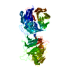
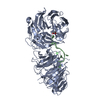
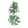
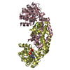
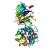
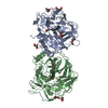
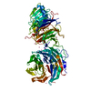
| Entry | Database: PDB / ID: 5gxi | ||||||
|---|---|---|---|---|---|---|---|
| Title | Structure of the Gemin5 WD40 domain in complex with AAUUUUUGAG | ||||||
 Components Components |
| ||||||
 Keywords Keywords | RNA BINDING PROTEIN/RNA /  snRNP assembly / snRNA / Structural Genomics / Structural Genomics Consortium / SGC / RNA BINDING PROTEIN-RNA complex snRNP assembly / snRNA / Structural Genomics / Structural Genomics Consortium / SGC / RNA BINDING PROTEIN-RNA complex | ||||||
| Function / homology |  Function and homology information Function and homology informationSMN-Gemin2 complex / Gemini of coiled bodies / SMN complex / U4atac snRNA binding / snRNA binding / RNA 7-methylguanosine cap binding / SMN-Sm protein complex / U4 snRNA binding / spliceosomal snRNP assembly / U1 snRNA binding ...SMN-Gemin2 complex / Gemini of coiled bodies / SMN complex / U4atac snRNA binding / snRNA binding / RNA 7-methylguanosine cap binding / SMN-Sm protein complex / U4 snRNA binding / spliceosomal snRNP assembly / U1 snRNA binding / mRNA 3'-UTR binding / mRNA splicing, via spliceosome / ribosome binding / regulation of translation / snRNP Assembly / SARS-CoV-2 modulates host translation machinery / protein-containing complex assembly / nuclear body / translation / RNA binding / nucleoplasm / membrane / nucleus / cytosol / cytoplasmSimilarity search - Function | ||||||
| Biological species |  Homo sapiens (human) Homo sapiens (human) | ||||||
| Method | X-RAY DIFFRACTION / MOLECULAR REPLACEMENT / Resolution: 1.85 Å | ||||||
 Authors Authors | Xu, C. / He, H. / Li, Y. / Dong, A. / Bountra, C. / Arrowsmith, C.H. / Edwards, A.M. / Min, J. / Structural Genomics Consortium (SGC) | ||||||
 Citation Citation | Journal: Genes Dev. / Year: 2016 Title: Structural insights into Gemin5-guided selection of pre-snRNAs for snRNP assembly Authors: Xu, C. / Ishikawa, H. / Izumikawa, K. / Li, L. / He, H. / Nobe, Y. / Yamauchi, Y. / Shahjee, H.M. / Wu, X.-H. / Yu, Y.-T. / Isobe, T. / Takahashi, N. / Min, J. | ||||||
| History |
|
- Structure visualization
| Structure viewer | Molecule: MolmilJmol/JSmol |
|---|
- Downloads & links
-Download
| PDBx/mmCIF format | 5gxi.cif.gz | 161.6 KB | Display | PDBx/mmCIF format |
|---|---|---|---|---|
| PDB format | pdb5gxi.ent.gz | 120.2 KB | Display | PDB format |
| PDBx/mmJSON format | 5gxi.json.gz | Tree view | PDBx/mmJSON format | |
| Others |  Other downloads Other downloads |
-Validation report
| Arichive directory | https://data.pdbj.org/pub/pdb/validation_reports/gx/5gxiftp://data.pdbj.org/pub/pdb/validation_reports/gx/5gxi | HTTPS FTP |
|---|
-Related structure data
| Related structure data | 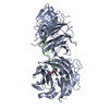 5gxhC  5teeC  5tefC  5thaC  2hesS 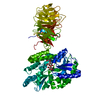 3dm0S  3ow8S C: citing same article ( S: Starting model for refinement |
|---|---|
| Similar structure data | |
| Other databases |


-Links
 PDBj
PDBj


- Assembly
| Deposited unit | 
| ||||||||
|---|---|---|---|---|---|---|---|---|---|
| 1 |
| ||||||||
| Unit cell |
|
-Components
| #1: Protein | / Gemin5 Mass: 84337.359 Da / Num. of mol.: 1 / Fragment: UNP residues 1-739 / Mutation: R682Q Source method: isolated from a genetically manipulated source Source: (gene. exp.) Homo sapiens (human) / Gene: GEMIN5 / Cell line (production host): Sf9 / Production host:   Spodoptera frugiperda (fall armyworm) / References: UniProt: Q8TEQ6 Spodoptera frugiperda (fall armyworm) / References: UniProt: Q8TEQ6 | ||
|---|---|---|---|
| #2: RNA chain | Mass: 3163.901 Da / Num. of mol.: 1 / Source method: obtained synthetically / Source: (synth.) Homo sapiens (human) | ||
| #3: Chemical | ChemComp-UNX /   Num. of mol.: 22 / Source method: obtained synthetically Num. of mol.: 22 / Source method: obtained synthetically#4: Water | ChemComp-HOH / | Water Mass: 18.015 Da / Num. of mol.: 381 / Source method: isolated from a natural source / Formula: H2O Mass: 18.015 Da / Num. of mol.: 381 / Source method: isolated from a natural source / Formula: H2O |
-Experimental details
-Experiment
| Experiment | Method: X-RAY DIFFRACTION / Number of used crystals: 1 |
|---|
- Sample preparation
| Crystal | Density Matthews: 2.23 Å3/Da / Density % sol: 44.77 % |
|---|---|
| Crystal grow | Temperature: 291 K / Method: vapor diffusion, sitting drop / pH: 6.5 Details: 0.1 M sodium citrate, 0.2 M ammonium acetate, 15% PEG 4000 |
-Data collection
| Diffraction | Mean temperature: 100 K |
|---|---|
| Diffraction source | Source: ROTATING ANODE / Type: RIGAKU FR-E DW / Wavelength: 1.5418 Å |
| Detector | Type: RIGAKU RAXIS II / Detector: IMAGE PLATE / Date: Apr 28, 2016 |
| Radiation | Protocol: SINGLE WAVELENGTH / Monochromatic (M) / Laue (L): M / Scattering type: x-ray |
| Radiation wavelength | Wavelength: 1.5418 Å / Relative weight: 1 |
| Reflection | Resolution: 1.85→28.92 Å / Num. obs: 65191 / % possible obs: 99.9 % / Redundancy: 7.3 % / CC1/2: 0.998 / Net I/σ(I): 14.9 |
| Reflection shell | Resolution: 1.85→1.9 Å / Redundancy: 7 % / Mean I/σ(I) obs: 1.8 / CC1/2: 0.592 / % possible all: 99.9 |
- Processing
| Software |
| ||||||||||||||||||||||||||||||||||||||||||||||||||||||||||||||||||||||||||||||||||||||||||||||||||||||||||||||||||||||||||||||||||||||||||||||||||||||||||||||||||||||||||||||||||||||
|---|---|---|---|---|---|---|---|---|---|---|---|---|---|---|---|---|---|---|---|---|---|---|---|---|---|---|---|---|---|---|---|---|---|---|---|---|---|---|---|---|---|---|---|---|---|---|---|---|---|---|---|---|---|---|---|---|---|---|---|---|---|---|---|---|---|---|---|---|---|---|---|---|---|---|---|---|---|---|---|---|---|---|---|---|---|---|---|---|---|---|---|---|---|---|---|---|---|---|---|---|---|---|---|---|---|---|---|---|---|---|---|---|---|---|---|---|---|---|---|---|---|---|---|---|---|---|---|---|---|---|---|---|---|---|---|---|---|---|---|---|---|---|---|---|---|---|---|---|---|---|---|---|---|---|---|---|---|---|---|---|---|---|---|---|---|---|---|---|---|---|---|---|---|---|---|---|---|---|---|---|---|---|---|
| Refinement | Method to determine structure: MOLECULAR REPLACEMENT Starting model: 3OW8,2HES,3DM0 Resolution: 1.85→28.9 Å / Cor.coef. Fo:Fc: 0.964 / Cor.coef. Fo:Fc free: 0.943 / SU B: 3.017 / SU ML: 0.088 / Cross valid method: THROUGHOUT / ESU R: 0.119 / ESU R Free: 0.119 / Details: HYDROGENS HAVE BEEN ADDED IN THE RIDING POSITIONS
| ||||||||||||||||||||||||||||||||||||||||||||||||||||||||||||||||||||||||||||||||||||||||||||||||||||||||||||||||||||||||||||||||||||||||||||||||||||||||||||||||||||||||||||||||||||||
| Solvent computation | Ion probe radii: 0.8 Å / Shrinkage radii: 0.8 Å / VDW probe radii: 1.2 Å | ||||||||||||||||||||||||||||||||||||||||||||||||||||||||||||||||||||||||||||||||||||||||||||||||||||||||||||||||||||||||||||||||||||||||||||||||||||||||||||||||||||||||||||||||||||||
| Displacement parameters | Biso mean: 24.115 Å2
| ||||||||||||||||||||||||||||||||||||||||||||||||||||||||||||||||||||||||||||||||||||||||||||||||||||||||||||||||||||||||||||||||||||||||||||||||||||||||||||||||||||||||||||||||||||||
| Refinement step | Cycle: 1 / Resolution: 1.85→28.9 Å
| ||||||||||||||||||||||||||||||||||||||||||||||||||||||||||||||||||||||||||||||||||||||||||||||||||||||||||||||||||||||||||||||||||||||||||||||||||||||||||||||||||||||||||||||||||||||
| Refine LS restraints |
|