 Movie
Movie Controller

[English] 日本語
 Yorodumi
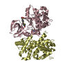
- PDB-5gx8: Crystal structure of solute-binding protein related to glycosamin... -
+ Open data

- Basic information
| Entry | Database: PDB / ID: 5gx8 | ||||||||||||
|---|---|---|---|---|---|---|---|---|---|---|---|---|---|
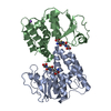
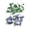
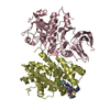
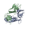
| Title | Crystal structure of solute-binding protein related to glycosaminoglycans | ||||||||||||
 Components Components | Extracellular solute-binding protein family 1 | ||||||||||||
 Keywords Keywords | SUGAR BINDING PROTEIN /  glycosaminoglycan / ABC transporter / solute-binding protein glycosaminoglycan / ABC transporter / solute-binding protein | ||||||||||||
| Function / homology |  Function and homology information Function and homology information | ||||||||||||
| Biological species |  Streptobacillus moniliformis DSM 12112 (bacteria) Streptobacillus moniliformis DSM 12112 (bacteria) | ||||||||||||
| Method | X-RAY DIFFRACTION / SYNCHROTRON / MOLECULAR REPLACEMENT / Resolution: 1.778 Å | ||||||||||||
 Authors Authors | Oiki, S. / Mikami, B. / Murata, K. / Hashimoto, W. | ||||||||||||
| Funding support |  Japan, 3items Japan, 3items
| ||||||||||||
 Citation Citation | Journal: Sci Rep / Year: 2017 Title: A bacterial ABC transporter enables import of mammalian host glycosaminoglycans Authors: Oiki, S. / Mikami, B. / Maruyama, Y. / Murata, K. / Hashimoto, W. | ||||||||||||
| History |
|
- Structure visualization
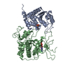
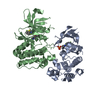
| Structure viewer | Molecule: MolmilJmol/JSmol |
|---|
- Downloads & links
-Download
| PDBx/mmCIF format | 5gx8.cif.gz | 221.1 KB | Display | PDBx/mmCIF format |
|---|---|---|---|---|
| PDB format | pdb5gx8.ent.gz | 174.7 KB | Display | PDB format |
| PDBx/mmJSON format | 5gx8.json.gz | Tree view | PDBx/mmJSON format | |
| Others |  Other downloads Other downloads |
-Validation report
| Arichive directory | https://data.pdbj.org/pub/pdb/validation_reports/gx/5gx8ftp://data.pdbj.org/pub/pdb/validation_reports/gx/5gx8 | HTTPS FTP |
|---|
-Related structure data
| Related structure data |  5gubSC 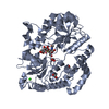 5gx6C  5gx7C C: citing same article ( S: Starting model for refinement |
|---|---|
| Similar structure data |
-Links
 PDBj
PDBj


- Assembly
| Deposited unit | 
| ||||||||
|---|---|---|---|---|---|---|---|---|---|
| 1 | 
| ||||||||
| 2 | 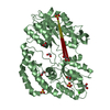
| ||||||||
| 3 |
| ||||||||
| Unit cell |
|
-Components
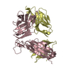
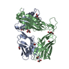
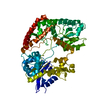
-Protein , 1 types, 2 molecules AB
| #1: Protein | Mass: 54271.309 Da / Num. of mol.: 2 / Fragment: UNP residues 28-500 Source method: isolated from a genetically manipulated source Source: (gene. exp.) Streptobacillus moniliformis DSM 12112 (bacteria)Strain: DSM 12112 / Gene: Smon_0123 / Production host: Escherichia coli (E. coli) / References: UniProt: D1AWE0 |
|---|
-Non-polymers , 5 types, 787 molecules 


| #2: Chemical |  Mass: 40.078 Da / Num. of mol.: 2 / Source method: obtained synthetically / Formula: Ca Mass: 40.078 Da / Num. of mol.: 2 / Source method: obtained synthetically / Formula: Ca#3: Chemical | ChemComp-EDO / Ethylene glycol Mass: 62.068 Da / Num. of mol.: 8 / Source method: obtained synthetically / Formula: C2H6O2 Mass: 62.068 Da / Num. of mol.: 8 / Source method: obtained synthetically / Formula: C2H6O2#4: Chemical | ChemComp-SO4 / Sulfate Mass: 96.063 Da / Num. of mol.: 7 / Source method: obtained synthetically / Formula: SO4 Mass: 96.063 Da / Num. of mol.: 7 / Source method: obtained synthetically / Formula: SO4#5: Chemical | ChemComp-TAR / | Tartaric acid Mass: 150.087 Da / Num. of mol.: 1 / Source method: obtained synthetically / Formula: C4H6O6 Mass: 150.087 Da / Num. of mol.: 1 / Source method: obtained synthetically / Formula: C4H6O6#6: Water | ChemComp-HOH / | WaterMass: 18.015 Da / Num. of mol.: 769 / Source method: isolated from a natural source / Formula: H2O |
|---|
-Experimental details
-Experiment
| Experiment | Method: X-RAY DIFFRACTION / Number of used crystals: 1 |
|---|
- Sample preparation
| Crystal | Density Matthews: 3.41 Å3/Da / Density % sol: 63.98 % |
|---|---|
| Crystal grow | Temperature: 293 K / Method: vapor diffusion, sitting drop Details: 0.2M potassium sodium tartrate tetrahydrate, 0.1M tri-sodium citrate dehydrate (pH 5.6), 2M ammonium sulfate |
-Data collection
| Diffraction | Mean temperature: 100 K |
|---|---|
| Diffraction source | Source: SYNCHROTRON / Site: SPring-8 / Beamline: BL38B1 / Wavelength: 1 Å |
| Detector | Type: RAYONIX MX225HE / Detector: CCD / Date: Dec 4, 2015 |
| Radiation | Protocol: SINGLE WAVELENGTH / Monochromatic (M) / Laue (L): M / Scattering type: x-ray |
| Radiation wavelength | Wavelength: 1 Å / Relative weight: 1 |
| Reflection | Resolution: 1.78→50 Å / Num. obs: 139193 / % possible obs: 98.6 % / Redundancy: 2 % / Rmerge(I) obs: 0.057 / Net I/σ(I): 29.2 |
| Reflection shell | Resolution: 1.78→1.81 Å / Rmerge(I) obs: 0.407 |
- Processing
| Software |
| |||||||||||||||||||||||||||||||||||||||||||||||||||||||||||||||||||||||||||||||||||||||||||||||||||||||||||||||||||||||||||||||||||||||||||||||||||||||||||||||||||||||||||||||||||||||||||||||||||||||||||||||||||||||||
|---|---|---|---|---|---|---|---|---|---|---|---|---|---|---|---|---|---|---|---|---|---|---|---|---|---|---|---|---|---|---|---|---|---|---|---|---|---|---|---|---|---|---|---|---|---|---|---|---|---|---|---|---|---|---|---|---|---|---|---|---|---|---|---|---|---|---|---|---|---|---|---|---|---|---|---|---|---|---|---|---|---|---|---|---|---|---|---|---|---|---|---|---|---|---|---|---|---|---|---|---|---|---|---|---|---|---|---|---|---|---|---|---|---|---|---|---|---|---|---|---|---|---|---|---|---|---|---|---|---|---|---|---|---|---|---|---|---|---|---|---|---|---|---|---|---|---|---|---|---|---|---|---|---|---|---|---|---|---|---|---|---|---|---|---|---|---|---|---|---|---|---|---|---|---|---|---|---|---|---|---|---|---|---|---|---|---|---|---|---|---|---|---|---|---|---|---|---|---|---|---|---|---|---|---|---|---|---|---|---|---|---|---|---|---|---|---|---|---|
| Refinement | Method to determine structure: MOLECULAR REPLACEMENT Starting model: 5GUB Resolution: 1.778→39.364 Å / SU ML: 0.2 / Cross valid method: THROUGHOUT / σ(F): 1.34 / Phase error: 23.86 / Stereochemistry target values: ML
| |||||||||||||||||||||||||||||||||||||||||||||||||||||||||||||||||||||||||||||||||||||||||||||||||||||||||||||||||||||||||||||||||||||||||||||||||||||||||||||||||||||||||||||||||||||||||||||||||||||||||||||||||||||||||
| Solvent computation | Shrinkage radii: 0.9 Å / VDW probe radii: 1.2 Å / Solvent model: FLAT BULK SOLVENT MODEL | |||||||||||||||||||||||||||||||||||||||||||||||||||||||||||||||||||||||||||||||||||||||||||||||||||||||||||||||||||||||||||||||||||||||||||||||||||||||||||||||||||||||||||||||||||||||||||||||||||||||||||||||||||||||||
| Refinement step | Cycle: LAST / Resolution: 1.778→39.364 Å
| |||||||||||||||||||||||||||||||||||||||||||||||||||||||||||||||||||||||||||||||||||||||||||||||||||||||||||||||||||||||||||||||||||||||||||||||||||||||||||||||||||||||||||||||||||||||||||||||||||||||||||||||||||||||||
| Refine LS restraints |
| |||||||||||||||||||||||||||||||||||||||||||||||||||||||||||||||||||||||||||||||||||||||||||||||||||||||||||||||||||||||||||||||||||||||||||||||||||||||||||||||||||||||||||||||||||||||||||||||||||||||||||||||||||||||||
| LS refinement shell |
|
