 Movie
Movie Controller

[English] 日本語
 Yorodumi
- PDB-5gv7: Structure of NADH-cytochrome b5 reductase refined with the multip... -
+ Open data

- Basic information
| Entry | Database: PDB / ID: 5gv7 | ||||||
|---|---|---|---|---|---|---|---|
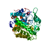
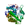
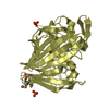
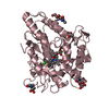
| Title | Structure of NADH-cytochrome b5 reductase refined with the multipolar atomic model at 0.80 A | ||||||
 Components Components | NADH-cytochrome b5 reductase 3 | ||||||
 Keywords Keywords |  OXIDOREDUCTASE / electron transfer / flavoprotein OXIDOREDUCTASE / electron transfer / flavoprotein | ||||||
| Function / homology |  Function and homology information Function and homology informationcytochrome-b5 reductase / cytochrome-b5 reductase activity, acting on NAD(P)H / cholesterol biosynthetic process / FAD binding / flavin adenine dinucleotide binding / mitochondrial outer membrane / endoplasmic reticulum membrane / mitochondrionSimilarity search - Function | ||||||
| Biological species |  Sus scrofa (pig) Sus scrofa (pig) | ||||||
| Method | X-RAY DIFFRACTION / SYNCHROTRON / MOLECULAR REPLACEMENT / Resolution: 0.8 Å | ||||||
 Authors Authors | Takaba, K. / Takeda, K. / Miki, K. | ||||||
 Citation Citation | Journal: Sci Rep / Year: 2017 Title: Distribution of valence electrons of the flavin cofactor in NADH-cytochrome b5 reductase. Authors: Takaba, K. / Takeda, K. / Kosugi, M. / Tamada, T. / Miki, K. | ||||||
| History |
|
- Structure visualization
| Structure viewer | Molecule: MolmilJmol/JSmol |
|---|
- Downloads & links
-Download
| PDBx/mmCIF format | 5gv7.cif.gz | 203.7 KB | Display | PDBx/mmCIF format |
|---|---|---|---|---|
| PDB format | pdb5gv7.ent.gz | 163.5 KB | Display | PDB format |
| PDBx/mmJSON format | 5gv7.json.gz | Tree view | PDBx/mmJSON format | |
| Others |  Other downloads Other downloads |
-Validation report
| Arichive directory | https://data.pdbj.org/pub/pdb/validation_reports/gv/5gv7ftp://data.pdbj.org/pub/pdb/validation_reports/gv/5gv7 | HTTPS FTP |
|---|
-Related structure data
| Related structure data | 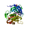 5gv8C 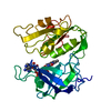 1ndhS C: citing same article ( S: Starting model for refinement |
|---|---|
| Similar structure data |


-Links
 PDBj
PDBj

- Assembly
| Deposited unit | 
| ||||||||
|---|---|---|---|---|---|---|---|---|---|
| 1 |
| ||||||||
| Unit cell |
|
-Components
| #1: Protein | Mass: 30873.674 Da / Num. of mol.: 1 Source method: isolated from a genetically manipulated source Source: (gene. exp.) Sus scrofa (pig) / Gene: CYB5R3, DIA1 / Production host:  Escherichia coli (E. coli) / References: UniProt: P83686, cytochrome-b5 reductase Escherichia coli (E. coli) / References: UniProt: P83686, cytochrome-b5 reductase | ||
|---|---|---|---|
| #2: Chemical | ChemComp-FAD / Flavin adenine dinucleotide  Mass: 785.550 Da / Num. of mol.: 1 / Source method: obtained synthetically / Formula: C27H33N9O15P2 / Comment: FAD*YM Mass: 785.550 Da / Num. of mol.: 1 / Source method: obtained synthetically / Formula: C27H33N9O15P2 / Comment: FAD*YM | ||
| #3: Chemical | ChemComp-GOL / Glycerol  Mass: 92.094 Da / Num. of mol.: 4 / Source method: obtained synthetically / Formula: C3H8O3 Mass: 92.094 Da / Num. of mol.: 4 / Source method: obtained synthetically / Formula: C3H8O3#4: Water | ChemComp-HOH / | Water Mass: 18.015 Da / Num. of mol.: 616 / Source method: isolated from a natural source / Formula: H2O Mass: 18.015 Da / Num. of mol.: 616 / Source method: isolated from a natural source / Formula: H2O |
-Experimental details
-Experiment
| Experiment | Method: X-RAY DIFFRACTION / Number of used crystals: 1 |
|---|
- Sample preparation
| Crystal | Density Matthews: 2.42 Å3/Da / Density % sol: 49.25 % |
|---|---|
| Crystal grow | Temperature: 293 K / Method: vapor diffusion / pH: 7.5 / Details: PEG 4000, DTT, K-phos buffer |
-Data collection
| Diffraction | Mean temperature: 40 K Ambient temp details: The author used a cryo-stream of helium. |
|---|---|
| Diffraction source | Source: SYNCHROTRON / Site: SPring-8  / Beamline: BL41XU / Wavelength: 0.65 Å / Beamline: BL41XU / Wavelength: 0.65 Å |
| Detector | Type: RAYONIX MX225HE / Detector: CCD / Date: Feb 2, 2011 |
| Radiation | Protocol: SINGLE WAVELENGTH / Monochromatic (M) / Laue (L): M / Scattering type: x-ray |
| Radiation wavelength | Wavelength: 0.65 Å / Relative weight: 1 |
| Reflection | Resolution: 0.8→29.3 Å / Num. obs: 314475 / % possible obs: 99.9 % / Redundancy: 7.5 % / Rsym value: 0.067 / Net I/σ(I): 31.6 |
| Reflection shell | Resolution: 0.8→0.81 Å / Redundancy: 6.8 % / Mean I/σ(I) obs: 1.3 / % possible all: 99.9 |
- Processing
| Software |
| ||||||||||||||||||||
|---|---|---|---|---|---|---|---|---|---|---|---|---|---|---|---|---|---|---|---|---|---|
| Refinement | Method to determine structure: MOLECULAR REPLACEMENT Starting model: 1NDH Resolution: 0.8→29.3 Å / Cross valid method: FREE R-VALUE Details: The model was finally refined with aspherical atomic model by using MOPRO program.
| ||||||||||||||||||||
| Refinement step | Cycle: LAST / Resolution: 0.8→29.3 Å
|
