 Movie
Movie Controller

[English] 日本語
 Yorodumi
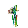
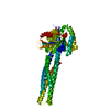
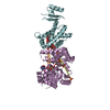
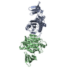
- PDB-5gns: Structures of human Mitofusin 1 provide insight into mitochondria... -
+ Open data

- Basic information
| Entry | Database: PDB / ID: 5gns | ||||||||||||
|---|---|---|---|---|---|---|---|---|---|---|---|---|---|
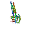
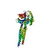
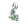
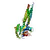
| Title | Structures of human Mitofusin 1 provide insight into mitochondrial tethering | ||||||||||||
 Components Components | Mitofusin-1 | ||||||||||||
 Keywords Keywords |  HYDROLASE / Mitofusin 1 / BDLP-like folding HYDROLASE / Mitofusin 1 / BDLP-like folding | ||||||||||||
| Function / homology |  Function and homology information Function and homology informationRHOT2 GTPase cycle / outer mitochondrial membrane protein complex / mitochondrion localization / GTP metabolic process / positive regulation of mitochondrial membrane potential / mitochondrial fusion / PINK1-PRKN Mediated Mitophagy / Hydrolases; Acting on acid anhydrides; Acting on GTP to facilitate cellular and subcellular movement / Factors involved in megakaryocyte development and platelet production / mitochondrial outer membrane ...RHOT2 GTPase cycle / outer mitochondrial membrane protein complex / mitochondrion localization / GTP metabolic process / positive regulation of mitochondrial membrane potential / mitochondrial fusion / PINK1-PRKN Mediated Mitophagy / Hydrolases; Acting on acid anhydrides; Acting on GTP to facilitate cellular and subcellular movement / Factors involved in megakaryocyte development and platelet production / mitochondrial outer membrane / GTPase activity / GTP binding / mitochondrion / membrane / identical protein bindingSimilarity search - Function | ||||||||||||
| Biological species |  Homo sapiens (human) Homo sapiens (human) | ||||||||||||
| Method | X-RAY DIFFRACTION / SYNCHROTRON / MOLECULAR REPLACEMENT / Resolution: 2.702 Å | ||||||||||||
 Authors Authors | Qi, Y. / Yan, L. / Yu, C. / Guo, X. / Zhou, X. / Hu, X. / Huang, X. / Rao, Z. / Lou, Z. / Hu, J. | ||||||||||||
| Funding support |  China, Armenia, 3items China, Armenia, 3items
| ||||||||||||
 Citation Citation | Journal: To Be Published Title: Structures of human Mitofusin 1 provide insight into mitochondrial tethering Authors: Qi, Y. / Yan, L. / Yu, C. / Guo, X. / Zhou, X. / Hu, X. / Huang, X. / Rao, Z. / Lou, Z. / Hu, J. | ||||||||||||
| History |
|
- Structure visualization
| Structure viewer | Molecule: MolmilJmol/JSmol |
|---|
- Downloads & links
-Download
| PDBx/mmCIF format | 5gns.cif.gz | 90.6 KB | Display | PDBx/mmCIF format |
|---|---|---|---|---|
| PDB format | pdb5gns.ent.gz | 67.7 KB | Display | PDB format |
| PDBx/mmJSON format | 5gns.json.gz | Tree view | PDBx/mmJSON format | |
| Others |  Other downloads Other downloads |
-Validation report
| Arichive directory | https://data.pdbj.org/pub/pdb/validation_reports/gn/5gnsftp://data.pdbj.org/pub/pdb/validation_reports/gn/5gns | HTTPS FTP |
|---|
-Related structure data
| Related structure data | |
|---|---|
| Similar structure data |


-Links
 PDBj
PDBj

- Assembly
| Deposited unit | 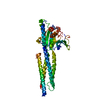
| ||||||||
|---|---|---|---|---|---|---|---|---|---|
| 1 |
| ||||||||
| Unit cell |
|
-Components
| #1: Protein | Mass: 47334.785 Da / Num. of mol.: 1 / Fragment: UNP residues 1-364 / Mutation: K88A Source method: isolated from a genetically manipulated source Source: (gene. exp.) Homo sapiens (human) / Gene: MFN1 / Production host:  Escherichia coli (E. coli) Escherichia coli (E. coli)References: UniProt: Q8IWA4, Hydrolases; Acting on acid anhydrides; Acting on GTP to facilitate cellular and subcellular movement |
|---|---|
| #2: Chemical | ChemComp-GTP / Guanosine triphosphate  Mass: 523.180 Da / Num. of mol.: 1 / Source method: obtained synthetically / Formula: C10H16N5O14P3 / Comment: GTP, energy-carrying molecule*YM Mass: 523.180 Da / Num. of mol.: 1 / Source method: obtained synthetically / Formula: C10H16N5O14P3 / Comment: GTP, energy-carrying molecule*YM |
| #3: Water | ChemComp-HOH / Water Mass: 18.015 Da / Num. of mol.: 8 / Source method: isolated from a natural source / Formula: H2O Mass: 18.015 Da / Num. of mol.: 8 / Source method: isolated from a natural source / Formula: H2O |
-Experimental details
-Experiment
| Experiment | Method: X-RAY DIFFRACTION / Number of used crystals: 1 |
|---|
- Sample preparation
| Crystal | Density Matthews: 2.68 Å3/Da / Density % sol: 54.07 % |
|---|---|
| Crystal grow | Temperature: 289 K / Method: vapor diffusion, hanging drop Details: 200mM sodium phosphate monobasic monohydrate, 20%(w/v) PEG3350 |
-Data collection
| Diffraction | Mean temperature: 100 K |
|---|---|
| Diffraction source | Source: SYNCHROTRON / Site: SSRF / Beamline: BL17U / Wavelength: 0.9785 Å |
| Detector | Type: ADSC QUANTUM 315r / Detector: CCD / Date: Jun 28, 2016 |
| Radiation | Protocol: SINGLE WAVELENGTH / Monochromatic (M) / Laue (L): M / Scattering type: x-ray |
| Radiation wavelength | Wavelength: 0.9785 Å / Relative weight: 1 |
| Reflection | Resolution: 2.3→50 Å / Num. obs: 122150 / % possible obs: 99.1 % / Redundancy: 14.1 % / Net I/σ(I): 10.2 |
- Processing
| Software |
| |||||||||||||||||||||||||||||||||||||||||||||||||||||||||||||||||||||||||||||
|---|---|---|---|---|---|---|---|---|---|---|---|---|---|---|---|---|---|---|---|---|---|---|---|---|---|---|---|---|---|---|---|---|---|---|---|---|---|---|---|---|---|---|---|---|---|---|---|---|---|---|---|---|---|---|---|---|---|---|---|---|---|---|---|---|---|---|---|---|---|---|---|---|---|---|---|---|---|---|
| Refinement | Method to determine structure: MOLECULAR REPLACEMENT / Resolution: 2.702→40.005 Å / SU ML: 0.37 / Cross valid method: NONE / σ(F): 1.34 / Phase error: 29.83 / Stereochemistry target values: ML
| |||||||||||||||||||||||||||||||||||||||||||||||||||||||||||||||||||||||||||||
| Solvent computation | Shrinkage radii: 0.9 Å / VDW probe radii: 1.11 Å / Solvent model: FLAT BULK SOLVENT MODEL | |||||||||||||||||||||||||||||||||||||||||||||||||||||||||||||||||||||||||||||
| Refinement step | Cycle: LAST / Resolution: 2.702→40.005 Å
| |||||||||||||||||||||||||||||||||||||||||||||||||||||||||||||||||||||||||||||
| Refine LS restraints |
| |||||||||||||||||||||||||||||||||||||||||||||||||||||||||||||||||||||||||||||
| LS refinement shell |
|
