 Movie
Movie Controller


+ Open data

- Basic information
| Entry | Database: PDB / ID: 5gnd | ||||||
|---|---|---|---|---|---|---|---|
| Title | Structure of Deg protease HhoA from Synechocystis sp. PCC 6803 | ||||||
 Components Components |
| ||||||
 Keywords Keywords |  HYDROLASE / Serine Protease HYDROLASE / Serine Protease | ||||||
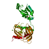
| Function / homology |  Function and homology information Function and homology informationouter membrane-bounded periplasmic space / serine-type endopeptidase activity / proteolysis / identical protein bindingSimilarity search - Function | ||||||
| Biological species |  Escherichia coli BL21 (bacteria) Escherichia coli BL21 (bacteria) | ||||||
| Method | X-RAY DIFFRACTION / SYNCHROTRON / MOLECULAR REPLACEMENT / Resolution: 2.5 Å | ||||||
 Authors Authors | Dong, W. / Wang, J. / Liu, L. | ||||||
 Citation Citation | Journal: Febs Lett. / Year: 2016 Title: Crystal structure of the zinc-bound HhoA protease from Synechocystis sp. PCC 6803 Authors: Dong, W. / Wang, J. / Niu, G. / Zhao, S. / Liu, L. | ||||||
| History |
|
- Structure visualization
| Structure viewer | Molecule: MolmilJmol/JSmol |
|---|
- Downloads & links
-Download
| PDBx/mmCIF format | 5gnd.cif.gz | 122.3 KB | Display | PDBx/mmCIF format |
|---|---|---|---|---|
| PDB format | pdb5gnd.ent.gz | 94 KB | Display | PDB format |
| PDBx/mmJSON format | 5gnd.json.gz | Tree view | PDBx/mmJSON format | |
| Others |  Other downloads Other downloads |
-Validation report
| Arichive directory | https://data.pdbj.org/pub/pdb/validation_reports/gn/5gndftp://data.pdbj.org/pub/pdb/validation_reports/gn/5gnd | HTTPS FTP |
|---|
-Related structure data
| Related structure data | 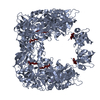 5b6lSC S: Starting model for refinement C: citing same article ( |
|---|---|
| Similar structure data |


-Links
 PDBj
PDBj
- Assembly
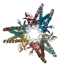
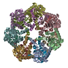
| Deposited unit | 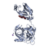
| ||||||||||||||||||
|---|---|---|---|---|---|---|---|---|---|---|---|---|---|---|---|---|---|---|---|
| 1 | 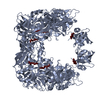
| ||||||||||||||||||
| Unit cell |
| ||||||||||||||||||
| Components on special symmetry positions |
|
-Components
| #1: Protein | Mass: 36868.691 Da / Num. of mol.: 1 / Fragment: UNP residues 56-394 Source method: isolated from a genetically manipulated source Source: (gene. exp.) Strain: PCC 6803 / Kazusa / Gene: hhoA / Plasmid: pET-22b(+) / Production host: Escherichia coli BL21(DE3) (bacteria) / Strain (production host): BL21(DE3) / References: UniProt: P72780 |
|---|---|
| #2: Protein/peptide | Mass: 544.644 Da / Num. of mol.: 1 / Source method: isolated from a natural source Details: This peptide is a co-purified peptide produced by Escherichia coli BL21(DE3). Source: (natural) Escherichia coli BL21(DE3) (bacteria) / Strain: BL21(DE3) |
| #3: Chemical | ChemComp-ZN /   Mass: 65.409 Da / Num. of mol.: 1 / Source method: obtained synthetically / Formula: Zn Mass: 65.409 Da / Num. of mol.: 1 / Source method: obtained synthetically / Formula: Zn |
| #4: Chemical | ChemComp-NA /   Mass: 22.990 Da / Num. of mol.: 1 / Source method: obtained synthetically / Formula: Na Mass: 22.990 Da / Num. of mol.: 1 / Source method: obtained synthetically / Formula: Na |
| #5: Water | ChemComp-HOH / Water Mass: 18.015 Da / Num. of mol.: 64 / Source method: isolated from a natural source / Formula: H2O Mass: 18.015 Da / Num. of mol.: 64 / Source method: isolated from a natural source / Formula: H2O |
-Experimental details
-Experiment
| Experiment | Method: X-RAY DIFFRACTION / Number of used crystals: 1 |
|---|
- Sample preparation
| Crystal | Density Matthews: 2.67 Å3/Da / Density % sol: 54.01 % |
|---|---|
| Crystal grow | Temperature: 289 K / Method: vapor diffusion, sitting drop / pH: 4.6 / Details: 0.1 M Na acetate, 25%(w/v) PEG4000 |
-Data collection
| Diffraction | Mean temperature: 100 K |
|---|---|
| Diffraction source | Source: SYNCHROTRON / Site: SSRF  / Beamline: BL17U / Wavelength: 0.9793 Å / Beamline: BL17U / Wavelength: 0.9793 Å |
| Detector | Type: ADSC QUANTUM 315r / Detector: CCD / Date: May 5, 2016 |
| Radiation | Protocol: SINGLE WAVELENGTH / Monochromatic (M) / Laue (L): M / Scattering type: x-ray |
| Radiation wavelength | Wavelength: 0.9793 Å / Relative weight: 1 |
| Reflection | Resolution: 2.5→50 Å / Num. obs: 13981 / % possible obs: 100 % / Redundancy: 11.1 % / Net I/σ(I): 59.5 |
| Reflection shell | Resolution: 2.5→2.59 Å / Redundancy: 10.6 % / Mean I/σ(I) obs: 2.9 / CC1/2: 0.807 / % possible all: 100 |
- Processing
| Software |
| ||||||||||||||||||||||||||||||||||||||||||
|---|---|---|---|---|---|---|---|---|---|---|---|---|---|---|---|---|---|---|---|---|---|---|---|---|---|---|---|---|---|---|---|---|---|---|---|---|---|---|---|---|---|---|---|
| Refinement | Method to determine structure: MOLECULAR REPLACEMENT Starting model: 5B6L Resolution: 2.5→37.92 Å / SU ML: 0.37 / Cross valid method: FREE R-VALUE / σ(F): 1.34 / Phase error: 31.72
| ||||||||||||||||||||||||||||||||||||||||||
| Solvent computation | Shrinkage radii: 0.9 Å / VDW probe radii: 1.11 Å | ||||||||||||||||||||||||||||||||||||||||||
| Displacement parameters | Biso max: 93.32 Å2 / Biso mean: 37.37 Å2 / Biso min: 10.17 Å2 | ||||||||||||||||||||||||||||||||||||||||||
| Refinement step | Cycle: final / Resolution: 2.5→37.92 Å
| ||||||||||||||||||||||||||||||||||||||||||
| Refine LS restraints |
| ||||||||||||||||||||||||||||||||||||||||||
| LS refinement shell | Refine-ID: X-RAY DIFFRACTION / Total num. of bins used: 5
| ||||||||||||||||||||||||||||||||||||||||||
| Refinement TLS params. | Method: refined / Origin x: -18.9689 Å / Origin y: 14.4422 Å / Origin z: 32.6502 Å
| ||||||||||||||||||||||||||||||||||||||||||
| Refinement TLS group |
|
