 Movie
Movie Controller

[English] 日本語
 Yorodumi
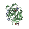
- PDB-5glk: Crystal structure of CoXyl43, GH43 beta-xylosidase/alpha-arabinof... -
+ Open data

- Basic information
| Entry | Database: PDB / ID: 5glk | ||||||
|---|---|---|---|---|---|---|---|
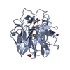
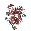
| Title | Crystal structure of CoXyl43, GH43 beta-xylosidase/alpha-arabinofuranosidase from a compost microbial metagenome, calcium-free form. | ||||||
 Components Components | Glycoside hydrolase family 43 | ||||||
 Keywords Keywords | HYDROLASE / Glycoside hydrolase family 43 | ||||||
| Function / homology |  Function and homology information Function and homology informationhydrolase activity, hydrolyzing O-glycosyl compounds / carbohydrate metabolic process Similarity search - Function | ||||||
| Biological species |  uncultured bacterium (environmental samples) uncultured bacterium (environmental samples) | ||||||
| Method | X-RAY DIFFRACTION / SYNCHROTRON / MOLECULAR REPLACEMENT / Resolution: 1.7 Å | ||||||
 Authors Authors | Matsuzawa, T. / Kishine, N. / Fujimoto, Z. / Yaoi, K. | ||||||
| Funding support |  Japan, 1items Japan, 1items
| ||||||
 Citation Citation | Journal: J. Biochem. / Year: 2017 Title: Crystal structure of metagenomic beta-xylosidase/ alpha-l-arabinofuranosidase activated by calcium. Authors: Matsuzawa, T. / Kaneko, S. / Kishine, N. / Fujimoto, Z. / Yaoi, K. #1: Journal: Appl. Microbiol. Biotechnol. / Year: 2015 Title: Screening, identification, and characterization of a GH43 family beta-xylosidase/alpha-arabinofuranosidase from a compost microbial metagenome. Authors: Matsuzawa, T. / Kaneko, S. / Yaoi, K. | ||||||
| History |
|
- Structure visualization
| Structure viewer | Molecule: MolmilJmol/JSmol |
|---|
- Downloads & links
-Download
| PDBx/mmCIF format | 5glk.cif.gz | 158.4 KB | Display | PDBx/mmCIF format |
|---|---|---|---|---|
| PDB format | pdb5glk.ent.gz | 120.8 KB | Display | PDB format |
| PDBx/mmJSON format | 5glk.json.gz | Tree view | PDBx/mmJSON format | |
| Others |  Other downloads Other downloads |
-Validation report
| Arichive directory | https://data.pdbj.org/pub/pdb/validation_reports/gl/5glkftp://data.pdbj.org/pub/pdb/validation_reports/gl/5glk | HTTPS FTP |
|---|
-Related structure data
| Related structure data |  5gllC 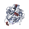 5glmC 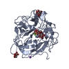 5glnC  5gloC 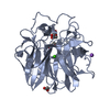 5glpC 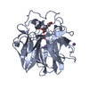 5glqC 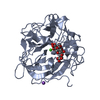 5glrC  4mlgS C: citing same article ( S: Starting model for refinement |
|---|---|
| Similar structure data |


-Links
 PDBj
PDBj


- Assembly
| Deposited unit | 
| ||||||||
|---|---|---|---|---|---|---|---|---|---|
| 1 | 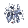
| ||||||||
| 2 | 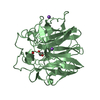
| ||||||||
| Unit cell |
|
-Components
| #1: Protein | / beta-xylosidase / alpha-arabinofuranosidase Mass: 38529.520 Da / Num. of mol.: 2 / Fragment: UNP RESIDUES 47-369 Source method: isolated from a genetically manipulated source Source: (gene. exp.) uncultured bacterium (environmental samples)Gene: coxyl43 / Plasmid: pET28b / Production host: Escherichia coli BL21(DE3) (bacteria) / Strain (production host): BL21(DE3) / References: UniProt: A0A0H5BL38#2: Chemical | ChemComp-NA /   Mass: 22.990 Da / Num. of mol.: 4 / Source method: obtained synthetically / Formula: Na Mass: 22.990 Da / Num. of mol.: 4 / Source method: obtained synthetically / Formula: Na#3: Chemical | Glycerol  Mass: 92.094 Da / Num. of mol.: 2 / Source method: obtained synthetically / Formula: C3H8O3 Mass: 92.094 Da / Num. of mol.: 2 / Source method: obtained synthetically / Formula: C3H8O3#4: Chemical | ChemComp-ACT / Acetate  Mass: 59.044 Da / Num. of mol.: 4 / Source method: obtained synthetically / Formula: C2H3O2 Mass: 59.044 Da / Num. of mol.: 4 / Source method: obtained synthetically / Formula: C2H3O2#5: Water | ChemComp-HOH / | Water Mass: 18.015 Da / Num. of mol.: 597 / Source method: isolated from a natural source / Formula: H2O Mass: 18.015 Da / Num. of mol.: 597 / Source method: isolated from a natural source / Formula: H2O |
|---|
-Experimental details
-Experiment
| Experiment | Method: X-RAY DIFFRACTION / Number of used crystals: 1 |
|---|
- Sample preparation
| Crystal | Density Matthews: 2.49 Å3/Da / Density % sol: 50.7 % / Description: Plate |
|---|---|
| Crystal grow | Temperature: 293 K / Method: vapor diffusion, sitting drop / pH: 5 / Details: 25% PEG3350, 8% Tacsimate |
-Data collection
| Diffraction | Mean temperature: 95 K |
|---|---|
| Diffraction source | Source: SYNCHROTRON / Site: Photon Factory / Beamline: AR-NW12A / Wavelength: 1 Å |
| Detector | Type: ADSC QUANTUM 210 / Detector: CCD / Date: May 24, 2015 |
| Radiation | Monochromator: Si III / Protocol: SINGLE WAVELENGTH / Monochromatic (M) / Laue (L): M / Scattering type: x-ray |
| Radiation wavelength | Wavelength: 1 Å / Relative weight: 1 |
| Reflection | Resolution: 1.7→100 Å / Num. obs: 78424 / % possible obs: 99.9 % / Redundancy: 14.7 % / Biso Wilson estimate: 12.349 Å2 / Rmerge(I) obs: 0.108 / Net I/σ(I): 26.3 |
| Reflection shell | Resolution: 1.7→1.76 Å / Redundancy: 14.1 % / Rmerge(I) obs: 0.883 / Mean I/σ(I) obs: 3.2 / % possible all: 100 |
- Processing
| Software |
| ||||||||||||||||||||||||||||||||||||||||||||||||||||||||||||||||||||||||||||||||||||||||||||||||||||||||||||||||||||||||||||||||||||||||||||||||||||||||||||||||||||||||||||||||||||||
|---|---|---|---|---|---|---|---|---|---|---|---|---|---|---|---|---|---|---|---|---|---|---|---|---|---|---|---|---|---|---|---|---|---|---|---|---|---|---|---|---|---|---|---|---|---|---|---|---|---|---|---|---|---|---|---|---|---|---|---|---|---|---|---|---|---|---|---|---|---|---|---|---|---|---|---|---|---|---|---|---|---|---|---|---|---|---|---|---|---|---|---|---|---|---|---|---|---|---|---|---|---|---|---|---|---|---|---|---|---|---|---|---|---|---|---|---|---|---|---|---|---|---|---|---|---|---|---|---|---|---|---|---|---|---|---|---|---|---|---|---|---|---|---|---|---|---|---|---|---|---|---|---|---|---|---|---|---|---|---|---|---|---|---|---|---|---|---|---|---|---|---|---|---|---|---|---|---|---|---|---|---|---|---|
| Refinement | Method to determine structure: MOLECULAR REPLACEMENT Starting model: 4MLG Resolution: 1.7→78.87 Å / Cor.coef. Fo:Fc: 0.966 / Cor.coef. Fo:Fc free: 0.954 / SU B: 1.926 / SU ML: 0.061 / Cross valid method: THROUGHOUT / ESU R: 0.093 / ESU R Free: 0.09 / Details: HYDROGENS HAVE BEEN ADDED IN THE RIDING POSITIONS
| ||||||||||||||||||||||||||||||||||||||||||||||||||||||||||||||||||||||||||||||||||||||||||||||||||||||||||||||||||||||||||||||||||||||||||||||||||||||||||||||||||||||||||||||||||||||
| Solvent computation | Ion probe radii: 0.8 Å / Shrinkage radii: 0.8 Å / VDW probe radii: 1.2 Å | ||||||||||||||||||||||||||||||||||||||||||||||||||||||||||||||||||||||||||||||||||||||||||||||||||||||||||||||||||||||||||||||||||||||||||||||||||||||||||||||||||||||||||||||||||||||
| Displacement parameters | Biso mean: 16.644 Å2
| ||||||||||||||||||||||||||||||||||||||||||||||||||||||||||||||||||||||||||||||||||||||||||||||||||||||||||||||||||||||||||||||||||||||||||||||||||||||||||||||||||||||||||||||||||||||
| Refinement step | Cycle: 1 / Resolution: 1.7→78.87 Å
| ||||||||||||||||||||||||||||||||||||||||||||||||||||||||||||||||||||||||||||||||||||||||||||||||||||||||||||||||||||||||||||||||||||||||||||||||||||||||||||||||||||||||||||||||||||||
| Refine LS restraints |
|