 Movie
Movie Controller


+ Open data

- Basic information
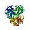
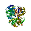
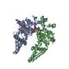
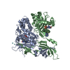
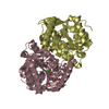
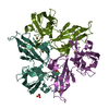
| Entry | Database: PDB / ID: 5gk2 | ||||||
|---|---|---|---|---|---|---|---|
| Title | The structure of the H302A mutant of StlD | ||||||
 Components Components | Ketosynthase StlD | ||||||
 Keywords Keywords |  TRANSFERASE / ketosynthase TRANSFERASE / ketosynthase | ||||||
| Function / homology | 3-Oxoacyl-[acyl-carrier-protein (ACP)] synthase III, C-terminal / 3-Oxoacyl-[acyl-carrier-protein (ACP)] synthase III C terminal / secondary metabolite biosynthetic process / acyltransferase activity / Thiolase-like / Photorhabdus luminescens subsp. laumondii TTO1 complete genome segment 8/17 Function and homology information Function and homology information | ||||||
| Biological species |  Photorhabdus luminescens subsp. laumondii (bacteria) Photorhabdus luminescens subsp. laumondii (bacteria) | ||||||
| Method | X-RAY DIFFRACTION / SYNCHROTRON / MOLECULAR REPLACEMENT / Resolution: 2.201 Å | ||||||
 Authors Authors | Mori, T. / Dngfeng, Y. / Morita, H. / Abe, I. | ||||||
 Citation Citation | Journal: Cell Chem Biol / Year: 2016 Title: Structural Insight into the Enzymatic Formation of Bacterial Stilbene. Authors: Mori, T. / Awakawa, T. / Shimomura, K. / Saito, Y. / Yang, D. / Morita, H. / Abe, I. | ||||||
| History |
|
- Structure visualization
| Structure viewer | Molecule: MolmilJmol/JSmol |
|---|
- Downloads & links
-Download
| PDBx/mmCIF format | 5gk2.cif.gz | 309.6 KB | Display | PDBx/mmCIF format |
|---|---|---|---|---|
| PDB format | pdb5gk2.ent.gz | 251 KB | Display | PDB format |
| PDBx/mmJSON format | 5gk2.json.gz | Tree view | PDBx/mmJSON format | |
| Others |  Other downloads Other downloads |
-Validation report
| Arichive directory | https://data.pdbj.org/pub/pdb/validation_reports/gk/5gk2ftp://data.pdbj.org/pub/pdb/validation_reports/gk/5gk2 | HTTPS FTP |
|---|
-Related structure data
| Related structure data | 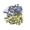 5gk0SC 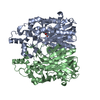 5gk1C S: Starting model for refinement C: citing same article ( |
|---|---|
| Similar structure data |


-Links
 PDBj
PDBj

- Assembly
| Deposited unit | 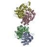
| ||||||||
|---|---|---|---|---|---|---|---|---|---|
| 1 | 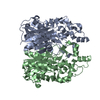
| ||||||||
| 2 | 
| ||||||||
| Unit cell |
|
-Components
| #1: Protein | Mass: 44684.375 Da / Num. of mol.: 4 / Mutation: H302A Source method: isolated from a genetically manipulated source Source: (gene. exp.) Photorhabdus luminescens subsp. laumondii (strain DSM 15139 / CIP 105565 / TT01) (bacteria)Strain: DSM 15139 / CIP 105565 / TT01 / Gene: plu2164 / Plasmid: pET28a / Production host: Escherichia coli (E. coli) / Strain (production host): BL21(DE3) codon plus / References: UniProt: Q7N4Z6#2: Water | ChemComp-HOH / | Water Mass: 18.015 Da / Num. of mol.: 568 / Source method: isolated from a natural source / Formula: H2O Mass: 18.015 Da / Num. of mol.: 568 / Source method: isolated from a natural source / Formula: H2O |
|---|
-Experimental details
-Experiment
| Experiment | Method: X-RAY DIFFRACTION / Number of used crystals: 1 |
|---|
- Sample preparation
| Crystal | Density Matthews: 2.44 Å3/Da / Density % sol: 49.68 % |
|---|---|
| Crystal grow | Temperature: 293 K / Method: vapor diffusion, sitting drop / Details: 12.5% PEG 4000, 100mM Tris-HCl (pH8.5) |
-Data collection
| Diffraction | Mean temperature: 100 K |
|---|---|
| Diffraction source | Source: SYNCHROTRON / Site: Photon Factory  / Beamline: AR-NE3A / Wavelength: 1 Å / Beamline: AR-NE3A / Wavelength: 1 Å |
| Detector | Type: DECTRIS PILATUS 2M-F / Detector: PIXEL / Date: Nov 16, 2015 |
| Radiation | Protocol: SINGLE WAVELENGTH / Monochromatic (M) / Laue (L): M / Scattering type: x-ray |
| Radiation wavelength | Wavelength: 1 Å / Relative weight: 1 |
| Reflection | Resolution: 2.2→50 Å / Num. obs: 86674 / % possible obs: 99.7 % / Redundancy: 3.45 % / Rmerge(I) obs: 0.079 / Net I/σ(I): 12.24 |
| Reflection shell | Resolution: 2.2→2.33 Å / Redundancy: 3.32 % / Rmerge(I) obs: 0.504 / Mean I/σ(I) obs: 2.38 / % possible all: 99.1 |
- Processing
| Software |
| |||||||||||||||||||||||||||||||||||||||||||||||||||||||||||||||||||||||||||||||||||||||||||||||||||||||||||||||||||||||||||||||||||||||||||||||||||||||||||||||||||||||||||||||||||||||||||||||||||||||||||||||||||||||||
|---|---|---|---|---|---|---|---|---|---|---|---|---|---|---|---|---|---|---|---|---|---|---|---|---|---|---|---|---|---|---|---|---|---|---|---|---|---|---|---|---|---|---|---|---|---|---|---|---|---|---|---|---|---|---|---|---|---|---|---|---|---|---|---|---|---|---|---|---|---|---|---|---|---|---|---|---|---|---|---|---|---|---|---|---|---|---|---|---|---|---|---|---|---|---|---|---|---|---|---|---|---|---|---|---|---|---|---|---|---|---|---|---|---|---|---|---|---|---|---|---|---|---|---|---|---|---|---|---|---|---|---|---|---|---|---|---|---|---|---|---|---|---|---|---|---|---|---|---|---|---|---|---|---|---|---|---|---|---|---|---|---|---|---|---|---|---|---|---|---|---|---|---|---|---|---|---|---|---|---|---|---|---|---|---|---|---|---|---|---|---|---|---|---|---|---|---|---|---|---|---|---|---|---|---|---|---|---|---|---|---|---|---|---|---|---|---|---|---|
| Refinement | Method to determine structure: MOLECULAR REPLACEMENT Starting model: 5GK0 Resolution: 2.201→41.698 Å / SU ML: 0.25 / Cross valid method: FREE R-VALUE / σ(F): 1.37 / Phase error: 23.85
| |||||||||||||||||||||||||||||||||||||||||||||||||||||||||||||||||||||||||||||||||||||||||||||||||||||||||||||||||||||||||||||||||||||||||||||||||||||||||||||||||||||||||||||||||||||||||||||||||||||||||||||||||||||||||
| Solvent computation | Shrinkage radii: 0.9 Å / VDW probe radii: 1.11 Å | |||||||||||||||||||||||||||||||||||||||||||||||||||||||||||||||||||||||||||||||||||||||||||||||||||||||||||||||||||||||||||||||||||||||||||||||||||||||||||||||||||||||||||||||||||||||||||||||||||||||||||||||||||||||||
| Refinement step | Cycle: LAST / Resolution: 2.201→41.698 Å
| |||||||||||||||||||||||||||||||||||||||||||||||||||||||||||||||||||||||||||||||||||||||||||||||||||||||||||||||||||||||||||||||||||||||||||||||||||||||||||||||||||||||||||||||||||||||||||||||||||||||||||||||||||||||||
| Refine LS restraints |
| |||||||||||||||||||||||||||||||||||||||||||||||||||||||||||||||||||||||||||||||||||||||||||||||||||||||||||||||||||||||||||||||||||||||||||||||||||||||||||||||||||||||||||||||||||||||||||||||||||||||||||||||||||||||||
| LS refinement shell |
|
