 Movie
Movie Controller

[English] 日本語
 Yorodumi
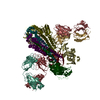
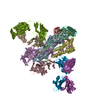
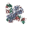
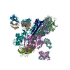
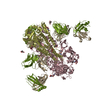
- PDB-5gjs: Crystal structure of H1 hemagglutinin from A/California/04/2009 i... -
+ Open data

- Basic information
| Entry | Database: PDB / ID: 5gjs | |||||||||
|---|---|---|---|---|---|---|---|---|---|---|
| Title | Crystal structure of H1 hemagglutinin from A/California/04/2009 in complex with a neutralizing antibody 3E1 | |||||||||
 Components Components |
| |||||||||
 Keywords Keywords |  IMMUNE SYSTEM / epitope / Fab / complementarity determining region / paratope IMMUNE SYSTEM / epitope / Fab / complementarity determining region / paratope | |||||||||
| Function / homology |  Function and homology informationviral budding from plasma membrane / clathrin-dependent endocytosis of virus by host cell / host cell surface receptor binding / apical plasma membrane / fusion of virus membrane with host plasma membrane / fusion of virus membrane with host endosome membrane / viral envelope / virion attachment to host cell / host cell plasma membrane / virion membrane Function and homology informationviral budding from plasma membrane / clathrin-dependent endocytosis of virus by host cell / host cell surface receptor binding / apical plasma membrane / fusion of virus membrane with host plasma membrane / fusion of virus membrane with host endosome membrane / viral envelope / virion attachment to host cell / host cell plasma membrane / virion membraneSimilarity search - Function | |||||||||
| Biological species |   Influenza A virus Influenza A virus Homo sapiens (human) Homo sapiens (human) | |||||||||
| Method | X-RAY DIFFRACTION / SYNCHROTRON / MOLECULAR REPLACEMENT / Resolution: 2.9 Å | |||||||||
 Authors Authors | Wang, W. / Zhang, T. / Ding, J. | |||||||||
 Citation Citation | Journal: Nat Commun / Year: 2016 Title: Human antibody 3E1 targets the HA stem region of H1N1 and H5N6 influenza A viruses Authors: Wang, W. / Sun, X. / Li, Y. / Su, J. / Ling, Z. / Zhang, T. / Wang, F. / Zhang, H. / Chen, H. / Ding, J. / Sun, B. | |||||||||
| History |
|
- Structure visualization
| Structure viewer | Molecule: MolmilJmol/JSmol |
|---|
- Downloads & links
-Download
| PDBx/mmCIF format | 5gjs.cif.gz | 353.8 KB | Display | PDBx/mmCIF format |
|---|---|---|---|---|
| PDB format | pdb5gjs.ent.gz | 286.8 KB | Display | PDB format |
| PDBx/mmJSON format | 5gjs.json.gz | Tree view | PDBx/mmJSON format | |
| Others |  Other downloads Other downloads |
-Validation report
| Arichive directory | https://data.pdbj.org/pub/pdb/validation_reports/gj/5gjsftp://data.pdbj.org/pub/pdb/validation_reports/gj/5gjs | HTTPS FTP |
|---|
-Related structure data
| Related structure data |  5gjtC  3lzjS 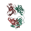 3nfsS C: citing same article ( S: Starting model for refinement |
|---|---|
| Similar structure data |
-Links
 PDBj
PDBj


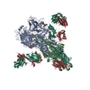
- Assembly
| Deposited unit | 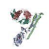
| ||||||||
|---|---|---|---|---|---|---|---|---|---|
| 1 | 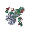
| ||||||||
| Unit cell |
|
-Components
-Protein , 2 types, 2 molecules AB
| #1: Protein | Mass: 36525.242 Da / Num. of mol.: 1 / Fragment: UNP residues 18-344 / Mutation: G212C, R227C Source method: isolated from a genetically manipulated source Source: (gene. exp.) Influenza A virus / Strain: swl A/California/04/2009 H1N1 / Gene: HA / Plasmid: pFast-HTB / Production host: Baculovirus expression vector pFastBac1-HM / Strain (production host): Sf9 / References: UniProt: C3W5S1 |
|---|---|
| #2: Protein | Mass: 20842.113 Da / Num. of mol.: 1 / Fragment: neutralizing antibody 3E1 Source method: isolated from a genetically manipulated source Source: (gene. exp.) Influenza A virus / Strain: swl A/California/04/2009 H1N1 / Gene: HA / Plasmid: pFast-HTB / Production host: Baculovirus expression vector pFastBac1-HM / Strain (production host): Sf9 / References: UniProt: C3W5S1 |
-Antibody , 2 types, 2 molecules LH
| #3: Antibody | Mass: 23483.084 Da / Num. of mol.: 1 Source method: isolated from a genetically manipulated source Source: (gene. exp.) Homo sapiens (human) / Plasmid: AbVec-hIgKappa2 / Production host: Cloning vector AbVec-hIgKappa (others) |
|---|---|
| #4: Antibody | Mass: 23663.646 Da / Num. of mol.: 1 Source method: isolated from a genetically manipulated source Source: (gene. exp.) Homo sapiens (human) / Plasmid: AbVec-hIgG1 / Production host: Cloning vector AbVec-hIgG1 (others) |
-Sugars , 2 types, 2 molecules 
| #5: Polysaccharide | 2-acetamido-2-deoxy-beta-D-glucopyranose-(1-4)-2-acetamido-2-deoxy-beta-D-glucopyranose / Mass: 424.401 Da / Num. of mol.: 1 / Source method: obtained synthetically |
|---|---|
| #6: Sugar | ChemComp-NAG / N-Acetylglucosamine Type: D-saccharide, beta linking / Mass: 221.208 Da / Num. of mol.: 1 / Source method: obtained synthetically / Formula: C8H15NO6 Type: D-saccharide, beta linking / Mass: 221.208 Da / Num. of mol.: 1 / Source method: obtained synthetically / Formula: C8H15NO6 |
-Experimental details
-Experiment
| Experiment | Method: X-RAY DIFFRACTION / Number of used crystals: 1 |
|---|
- Sample preparation
| Crystal | Density Matthews: 2.85 Å3/Da / Density % sol: 56.89 % Description: THE ENTRY CONTAINS FRIEDEL PAIRS IN F_PLUS/MINUS COLUMNS. |
|---|---|
| Crystal grow | Temperature: 293 K / Method: vapor diffusion, hanging drop / pH: 6.5 / Details: 0.1M MES and 16% PEG 550MME |
-Data collection
| Diffraction | Mean temperature: 100 K |
|---|---|
| Diffraction source | Source: SYNCHROTRON / Site: SSRF  / Beamline: BL17U / Wavelength: 0.97914 Å / Beamline: BL17U / Wavelength: 0.97914 Å |
| Detector | Type: ADSC QUANTUM 315r / Detector: CCD / Date: Sep 19, 2014 |
| Radiation | Monochromator: GRAPHITE / Protocol: SINGLE WAVELENGTH / Monochromatic (M) / Laue (L): M / Scattering type: x-ray |
| Radiation wavelength | Wavelength: 0.97914 Å / Relative weight: 1 |
| Reflection | Resolution: 2.9→50 Å / Num. obs: 25201 / % possible obs: 93.8 % / Redundancy: 6.5 % / Biso Wilson estimate: 46.6 Å2 / Rmerge(I) obs: 0.16 / Net I/σ(I): 12.9 |
| Reflection shell | Resolution: 2.9→3 Å / Redundancy: 6.3 % / Rmerge(I) obs: 0.8 / Mean I/σ(I) obs: 2.6 / % possible all: 100 |
- Processing
| Software |
| |||||||||||||||||||||||||||||||||||||||||||||||||||||||||||||||||||||||||||||||||||||||||||||||||||||||||||||||||||||||||||||
|---|---|---|---|---|---|---|---|---|---|---|---|---|---|---|---|---|---|---|---|---|---|---|---|---|---|---|---|---|---|---|---|---|---|---|---|---|---|---|---|---|---|---|---|---|---|---|---|---|---|---|---|---|---|---|---|---|---|---|---|---|---|---|---|---|---|---|---|---|---|---|---|---|---|---|---|---|---|---|---|---|---|---|---|---|---|---|---|---|---|---|---|---|---|---|---|---|---|---|---|---|---|---|---|---|---|---|---|---|---|---|---|---|---|---|---|---|---|---|---|---|---|---|---|---|---|---|
| Refinement | Method to determine structure: MOLECULAR REPLACEMENT Starting model: 3LZJ, 3NFS Resolution: 2.9→47.98 Å / Cor.coef. Fo:Fc: 0.87 / Cor.coef. Fo:Fc free: 0.814 / SU B: 48.675 / SU ML: 0.411 / Cross valid method: THROUGHOUT / σ(F): 0 / ESU R Free: 0.502 / Details: U VALUES : WITH TLS ADDED,
| |||||||||||||||||||||||||||||||||||||||||||||||||||||||||||||||||||||||||||||||||||||||||||||||||||||||||||||||||||||||||||||
| Solvent computation | Ion probe radii: 0.8 Å / Shrinkage radii: 0.8 Å / VDW probe radii: 1.2 Å | |||||||||||||||||||||||||||||||||||||||||||||||||||||||||||||||||||||||||||||||||||||||||||||||||||||||||||||||||||||||||||||
| Displacement parameters | Biso max: 138.37 Å2 / Biso mean: 51.878 Å2 / Biso min: 20.67 Å2
| |||||||||||||||||||||||||||||||||||||||||||||||||||||||||||||||||||||||||||||||||||||||||||||||||||||||||||||||||||||||||||||
| Refinement step | Cycle: final / Resolution: 2.9→47.98 Å
| |||||||||||||||||||||||||||||||||||||||||||||||||||||||||||||||||||||||||||||||||||||||||||||||||||||||||||||||||||||||||||||
| Refine LS restraints |
| |||||||||||||||||||||||||||||||||||||||||||||||||||||||||||||||||||||||||||||||||||||||||||||||||||||||||||||||||||||||||||||
| LS refinement shell | Resolution: 2.901→2.977 Å / Total num. of bins used: 20
| |||||||||||||||||||||||||||||||||||||||||||||||||||||||||||||||||||||||||||||||||||||||||||||||||||||||||||||||||||||||||||||
| Refinement TLS params. | Method: refined / Refine-ID: X-RAY DIFFRACTION
| |||||||||||||||||||||||||||||||||||||||||||||||||||||||||||||||||||||||||||||||||||||||||||||||||||||||||||||||||||||||||||||
| Refinement TLS group |
|
