 Movie
Movie Controller

[English] 日本語
 Yorodumi
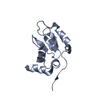
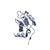
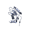
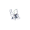
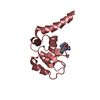
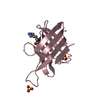
- PDB-5ghl: Crystal structure Analysis of the starch-binding domain of glucoa... -
+ Open data

- Basic information
| Entry | Database: PDB / ID: 5ghl | ||||||
|---|---|---|---|---|---|---|---|
| Title | Crystal structure Analysis of the starch-binding domain of glucoamylase from Aspergillus niger | ||||||
 Components Components | Glucoamylase Glucan 1,4-a-glucosidase Glucan 1,4-a-glucosidase | ||||||
 Keywords Keywords | HYDROLASE / beta-sheet structure | ||||||
| Function / homology |  Function and homology information Function and homology informationpolysaccharide metabolic process / glucan 1,4-alpha-glucosidase / glucan 1,4-alpha-glucosidase activity / starch binding / fungal-type vacuole / polysaccharide catabolic process / endoplasmic reticulumSimilarity search - Function | ||||||
| Biological species |  Aspergillus niger (mold) Aspergillus niger (mold) | ||||||
| Method | X-RAY DIFFRACTION / SYNCHROTRON / MOLECULAR REPLACEMENT / Resolution: 2 Å | ||||||
 Authors Authors | Miyake, H. / Suyama, Y. / Muraki, N. / Kusunoki, M. / Tanaka, A. | ||||||
 Citation Citation | Journal: Acta Crystallogr.,Sect.F / Year: 2017 Title: Crystal structure of the starch-binding domain of glucoamylase from Aspergillus niger. Authors: Suyama, Y. / Muraki, N. / Kusunoki, M. / Miyake, H. | ||||||
| History |
|
- Structure visualization
| Structure viewer | Molecule: MolmilJmol/JSmol |
|---|
- Downloads & links
-Download
| PDBx/mmCIF format | 5ghl.cif.gz | 101.4 KB | Display | PDBx/mmCIF format |
|---|---|---|---|---|
| PDB format | pdb5ghl.ent.gz | 78.2 KB | Display | PDB format |
| PDBx/mmJSON format | 5ghl.json.gz | Tree view | PDBx/mmJSON format | |
| Others |  Other downloads Other downloads |
-Validation report
| Arichive directory | https://data.pdbj.org/pub/pdb/validation_reports/gh/5ghlftp://data.pdbj.org/pub/pdb/validation_reports/gh/5ghl | HTTPS FTP |
|---|
-Related structure data
| Related structure data | 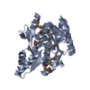 3nyiS S: Starting model for refinement |
|---|---|
| Similar structure data |

-Links
 PDBj
PDBj
- Assembly
| Deposited unit | 
| ||||||||
|---|---|---|---|---|---|---|---|---|---|
| 1 | 
| ||||||||
| 2 | 
| ||||||||
| 3 | 
| ||||||||
| 4 | 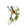
| ||||||||
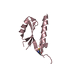
| Unit cell |
|
-Components
| #1: Protein | Glucan 1,4-a-glucosidase / 1 / 4-alpha-D-glucan glucohydrolase / Glucan 1 / 4-alpha-glucosidase Mass: 11884.820 Da / Num. of mol.: 4 Fragment: UNP residues 533-640, starch-binding domain of glucoamylase from Aspergillus niger Source method: isolated from a genetically manipulated source Source: (gene. exp.) Aspergillus niger (mold) / Gene: GLAA / Plasmid: pQE30 / Production host:  Escherichia coli (E. coli) / Strain (production host): M15 / References: UniProt: P69328, glucan 1,4-alpha-glucosidase Escherichia coli (E. coli) / Strain (production host): M15 / References: UniProt: P69328, glucan 1,4-alpha-glucosidase#2: Chemical | ChemComp-GOL / Glycerol  Mass: 92.094 Da / Num. of mol.: 4 / Source method: obtained synthetically / Formula: C3H8O3 Mass: 92.094 Da / Num. of mol.: 4 / Source method: obtained synthetically / Formula: C3H8O3#3: Chemical | ChemComp-SO4 / Sulfate  Mass: 96.063 Da / Num. of mol.: 4 / Source method: obtained synthetically / Formula: SO4 Mass: 96.063 Da / Num. of mol.: 4 / Source method: obtained synthetically / Formula: SO4#4: Water | ChemComp-HOH / | Water Mass: 18.015 Da / Num. of mol.: 204 / Source method: isolated from a natural source / Formula: H2O Mass: 18.015 Da / Num. of mol.: 204 / Source method: isolated from a natural source / Formula: H2O |
|---|
-Experimental details
-Experiment
| Experiment | Method: X-RAY DIFFRACTION / Number of used crystals: 1 |
|---|
- Sample preparation
| Crystal | Density Matthews: 2.72 Å3/Da / Density % sol: 54.75 % |
|---|---|
| Crystal grow | Temperature: 293 K / Method: vapor diffusion, hanging drop / pH: 7.5 / Details: Ammonium sulfate, PEG400, Glycerol |
-Data collection
| Diffraction | Mean temperature: 100 K | |||||||||||||||||||||||||||||||||||||||||||||||||||||||||||||||||||||||||||||||||||||||||||||||||||||||||||||||||||||||||||||||||||||||||||||||||||
|---|---|---|---|---|---|---|---|---|---|---|---|---|---|---|---|---|---|---|---|---|---|---|---|---|---|---|---|---|---|---|---|---|---|---|---|---|---|---|---|---|---|---|---|---|---|---|---|---|---|---|---|---|---|---|---|---|---|---|---|---|---|---|---|---|---|---|---|---|---|---|---|---|---|---|---|---|---|---|---|---|---|---|---|---|---|---|---|---|---|---|---|---|---|---|---|---|---|---|---|---|---|---|---|---|---|---|---|---|---|---|---|---|---|---|---|---|---|---|---|---|---|---|---|---|---|---|---|---|---|---|---|---|---|---|---|---|---|---|---|---|---|---|---|---|---|---|---|---|
| Diffraction source | Source: SYNCHROTRON / Site: Photon Factory  / Beamline: AR-NW12A / Wavelength: 0.978 Å / Beamline: AR-NW12A / Wavelength: 0.978 Å | |||||||||||||||||||||||||||||||||||||||||||||||||||||||||||||||||||||||||||||||||||||||||||||||||||||||||||||||||||||||||||||||||||||||||||||||||||
| Detector | Type: ADSC QUANTUM 210 / Detector: CCD / Date: Nov 27, 2004 | |||||||||||||||||||||||||||||||||||||||||||||||||||||||||||||||||||||||||||||||||||||||||||||||||||||||||||||||||||||||||||||||||||||||||||||||||||
| Radiation | Protocol: SINGLE WAVELENGTH / Monochromatic (M) / Laue (L): M / Scattering type: x-ray | |||||||||||||||||||||||||||||||||||||||||||||||||||||||||||||||||||||||||||||||||||||||||||||||||||||||||||||||||||||||||||||||||||||||||||||||||||
| Radiation wavelength | Wavelength: 0.978 Å / Relative weight: 1 | |||||||||||||||||||||||||||||||||||||||||||||||||||||||||||||||||||||||||||||||||||||||||||||||||||||||||||||||||||||||||||||||||||||||||||||||||||
| Reflection | Resolution: 2→50 Å / Num. obs: 34419 / % possible obs: 99.9 % / Redundancy: 8.5 % / Rmerge(I) obs: 0.107 / Χ2: 20.38 / Net I/σ(I): 44.5 / Num. measured all: 291560 | |||||||||||||||||||||||||||||||||||||||||||||||||||||||||||||||||||||||||||||||||||||||||||||||||||||||||||||||||||||||||||||||||||||||||||||||||||
| Reflection shell |
|
- Processing
| Software |
| ||||||||||||||||||||||||||||||||||||||||||||||||||||||||||||
|---|---|---|---|---|---|---|---|---|---|---|---|---|---|---|---|---|---|---|---|---|---|---|---|---|---|---|---|---|---|---|---|---|---|---|---|---|---|---|---|---|---|---|---|---|---|---|---|---|---|---|---|---|---|---|---|---|---|---|---|---|---|
| Refinement | Method to determine structure: MOLECULAR REPLACEMENT Starting model: 3NYI Resolution: 2→45.98 Å / Cor.coef. Fo:Fc: 0.953 / Cor.coef. Fo:Fc free: 0.919 / SU B: 4.052 / SU ML: 0.114 / Cross valid method: THROUGHOUT / σ(F): 0 / ESU R: 0.161 / ESU R Free: 0.155 / Stereochemistry target values: MAXIMUM LIKELIHOOD Details: HYDROGENS HAVE BEEN ADDED IN THE RIDING POSITIONS U VALUES : REFINED INDIVIDUALLY
| ||||||||||||||||||||||||||||||||||||||||||||||||||||||||||||
| Solvent computation | Ion probe radii: 0.8 Å / Shrinkage radii: 0.8 Å / VDW probe radii: 1.2 Å / Solvent model: MASK | ||||||||||||||||||||||||||||||||||||||||||||||||||||||||||||
| Displacement parameters | Biso max: 89.6 Å2 / Biso mean: 25.954 Å2 / Biso min: 7.83 Å2
| ||||||||||||||||||||||||||||||||||||||||||||||||||||||||||||
| Refinement step | Cycle: final / Resolution: 2→45.98 Å
| ||||||||||||||||||||||||||||||||||||||||||||||||||||||||||||
| Refine LS restraints |
| ||||||||||||||||||||||||||||||||||||||||||||||||||||||||||||
| LS refinement shell | Resolution: 2→2.052 Å / Rfactor Rfree error: 0 / Total num. of bins used: 20
|
