 Movie
Movie Controller

[English] 日本語
 Yorodumi
- PDB-5g5x: CBS domain tandem of site-2 protease from Archaeoglobus fulgidus ... -
+ Open data

- Basic information
| Entry | Database: PDB / ID: 5g5x | ||||||
|---|---|---|---|---|---|---|---|
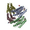
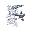
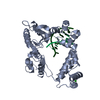
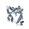
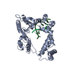
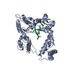
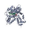
| Title | CBS domain tandem of site-2 protease from Archaeoglobus fulgidus in complex with llama Nanobody - nucleotide-bound form | ||||||
 Components Components |
| ||||||
 Keywords Keywords |  HYDROLASE / METALLOPROTEASE / SITE-2 PROTEASE / REGULATORY DOMAIN / NUCLEOTIDE-BINDING / CBS DOMAIN / CAMELID ANTIBODY / NANOBODY HYDROLASE / METALLOPROTEASE / SITE-2 PROTEASE / REGULATORY DOMAIN / NUCLEOTIDE-BINDING / CBS DOMAIN / CAMELID ANTIBODY / NANOBODY | ||||||
| Function / homology |  Function and homology informationmetalloendopeptidase activity / proteolysis / metal ion binding / plasma membrane Function and homology informationmetalloendopeptidase activity / proteolysis / metal ion binding / plasma membraneSimilarity search - Function | ||||||
| Biological species |   ARCHAEOGLOBUS FULGIDUS (archaea) ARCHAEOGLOBUS FULGIDUS (archaea) LAMA GLAMA (llama) LAMA GLAMA (llama) | ||||||
| Method | X-RAY DIFFRACTION / SYNCHROTRON / MOLECULAR REPLACEMENT / Resolution: 2.8 Å | ||||||
 Authors Authors | Schacherl, M. / Baumann, U. | ||||||
 Citation Citation | Journal: Biochim. Biophys. Acta / Year: 2017 Title: Crystallographic and biochemical characterization of the dimeric architecture of site-2 protease. Authors: Schacherl, M. / Gompert, M. / Pardon, E. / Lamkemeyer, T. / Steyaert, J. / Baumann, U. | ||||||
| History |
|
- Structure visualization
| Structure viewer | Molecule: MolmilJmol/JSmol |
|---|
- Downloads & links
-Download
| PDBx/mmCIF format | 5g5x.cif.gz | 109.9 KB | Display | PDBx/mmCIF format |
|---|---|---|---|---|
| PDB format | pdb5g5x.ent.gz | 84.6 KB | Display | PDB format |
| PDBx/mmJSON format | 5g5x.json.gz | Tree view | PDBx/mmJSON format | |
| Others |  Other downloads Other downloads |
-Validation report
| Arichive directory | https://data.pdbj.org/pub/pdb/validation_reports/g5/5g5xftp://data.pdbj.org/pub/pdb/validation_reports/g5/5g5x | HTTPS FTP |
|---|
-Related structure data
| Related structure data |  5g5rSC S: Starting model for refinement C: citing same article ( |
|---|---|
| Similar structure data |


-Links
 PDBj
PDBj


- Assembly
| Deposited unit | 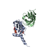
| ||||||||
|---|---|---|---|---|---|---|---|---|---|
| 1 | 
| ||||||||
| Unit cell |
|
-Components
| #1: Protein | Membrane-bound transcription factor site-2 protease Mass: 15469.021 Da / Num. of mol.: 1 / Fragment: REGULATORY DOMAIN, RESIDUES 236-362 Source method: isolated from a genetically manipulated source Details: C-TERMINAL STREPII-TAG / Source: (gene. exp.) ARCHAEOGLOBUS FULGIDUS (archaea) / Description: DSM 4304 GENOMIC DNA / Production host:  ESCHERICHIA COLI (E. coli) / Strain (production host): BL21(DE3) / Variant (production host): PLYSS / References: UniProt: O29915, EC: 3.4.24.85 ESCHERICHIA COLI (E. coli) / Strain (production host): BL21(DE3) / Variant (production host): PLYSS / References: UniProt: O29915, EC: 3.4.24.85 |
|---|---|
| #2: Antibody | Single-domain antibody Mass: 13318.636 Da / Num. of mol.: 1 Source method: isolated from a genetically manipulated source Details: NB330, SELECTED FROM LLAMA, C-TERMINAL 6XHIS- TAG / Source: (gene. exp.) LAMA GLAMA (llama) / Plasmid: PHEN6 / Production host: ESCHERICHIA COLI (E. coli) / Strain (production host): K-12 / Variant (production host): WK6 |
| #3: Chemical | ChemComp-AMP / Adenosine monophosphate  Mass: 347.221 Da / Num. of mol.: 1 / Source method: obtained synthetically / Formula: C10H14N5O7P / Comment: AMP*YM Mass: 347.221 Da / Num. of mol.: 1 / Source method: obtained synthetically / Formula: C10H14N5O7P / Comment: AMP*YM |
| #4: Chemical | ChemComp-ATP / Adenosine triphosphate  Mass: 507.181 Da / Num. of mol.: 1 / Source method: obtained synthetically / Formula: C10H16N5O13P3 / Comment: ATP, energy-carrying molecule*YM Mass: 507.181 Da / Num. of mol.: 1 / Source method: obtained synthetically / Formula: C10H16N5O13P3 / Comment: ATP, energy-carrying molecule*YM |
| Sequence details | D236-S362 |
-Experimental details
-Experiment
| Experiment | Method: X-RAY DIFFRACTION / Number of used crystals: 1 |
|---|
- Sample preparation
| Crystal | Density Matthews: 2.53 Å3/Da / Density % sol: 51.4 % / Description: NONE |
|---|---|
| Crystal grow | pH: 7.5 / Details: 0.1 M IMIDAZOLE PH 7.25, 0.2 M MGCL2, 12 % ETHANOL |
-Data collection
| Diffraction | Mean temperature: 293 K |
|---|---|
| Diffraction source | Source: SYNCHROTRON / Site: SLS  / Beamline: X06SA / Wavelength: 1 / Beamline: X06SA / Wavelength: 1 |
| Detector | Type: DECTRIS PILATUS 6M / Detector: PIXEL / Date: Dec 14, 2014 |
| Radiation | Protocol: SINGLE WAVELENGTH / Monochromatic (M) / Laue (L): M / Scattering type: x-ray |
| Radiation wavelength | Wavelength: 1 Å / Relative weight: 1 |
| Reflection | Resolution: 2.8→61.41 Å / Num. obs: 7966 / % possible obs: 99.7 % / Observed criterion σ(I): 2 / Redundancy: 9.58 % / Biso Wilson estimate: 72.63 Å2 / Rmerge(I) obs: 0.11 / Net I/σ(I): 14.21 |
| Reflection shell | Resolution: 2.8→2.97 Å / Redundancy: 9.27 % / Rmerge(I) obs: 0.89 / Mean I/σ(I) obs: 2.16 / % possible all: 99.3 |
- Processing
| Software |
| ||||||||||||||||||||||||||||
|---|---|---|---|---|---|---|---|---|---|---|---|---|---|---|---|---|---|---|---|---|---|---|---|---|---|---|---|---|---|
| Refinement | Method to determine structure: MOLECULAR REPLACEMENT Starting model: PDB ENTRY 5G5R Resolution: 2.8→52.433 Å / SU ML: 0.39 / σ(F): 1.36 / Phase error: 31.71 / Stereochemistry target values: ML
| ||||||||||||||||||||||||||||
| Solvent computation | Shrinkage radii: 0.9 Å / VDW probe radii: 1.11 Å / Solvent model: FLAT BULK SOLVENT MODEL / Bsol: 61.8 Å2 / ksol: 0.34 e/Å3 | ||||||||||||||||||||||||||||
| Displacement parameters | Biso mean: 87 Å2 | ||||||||||||||||||||||||||||
| Refinement step | Cycle: LAST / Resolution: 2.8→52.433 Å
| ||||||||||||||||||||||||||||
| Refine LS restraints |
| ||||||||||||||||||||||||||||
| LS refinement shell |
|
