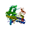
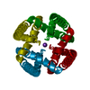
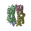
[English] 日本語
Yorodumi
- PDB-5fig: APO-CSP3 (COPPER STORAGE PROTEIN 3) FROM BACILLUS SUBTILIS -

+
Open data


ID or keywords:

Loading...

-
Basic information

Entry
Database: PDB / ID: 5fig
TitleAPO-CSP3 (COPPER STORAGE PROTEIN 3) FROM BACILLUS SUBTILIS
ComponentsCSP3
KeywordsCOPPER-BINDING PROTEIN / COPPER STORAGE / BACILLUS SUBTILIS
Function / homologySubstrate Binding Domain Of Dnak; Chain:A; Domain 2 - #360 / Protein of unknown function DUF326 / Domain of Unknown Function (DUF326) / Uncharacterized cysteine-rich protein YhjQ-like / Substrate Binding Domain Of Dnak; Chain:A; Domain 2 / Up-down Bundle / Mainly Alpha / Uncharacterized cysteine-rich protein YhjQ
Function and homology information
Biological speciesBACILLUS SUBTILIS (bacteria)
MethodX-RAY DIFFRACTION / SYNCHROTRON / MOLECULAR REPLACEMENT / Resolution: 1.7 Å
AuthorsVita, N. / Landolfi, G. / Basle, A. / Platsaki, S. / Waldron, K. / Dennison, C.
CitationJournal: Sci Rep / Year: 2016
Title: Bacterial cytosolic proteins with a high capacity for Cu(I) that protect against copper toxicity.
Authors: Vita, N. / Landolfi, G. / Basle, A. / Platsaki, S. / Lee, J. / Waldron, K.J. / Dennison, C.
History
DepositionSep 25, 2015Deposition site: PDBE / Processing site: PDBE
Revision 1.0Oct 12, 2016Provider: repository / Type: Initial release
Revision 1.1Apr 25, 2018Group: Data collection / Database references / Category: citation / diffrn_source
Item: _citation.country / _citation.journal_abbrev ..._citation.country / _citation.journal_abbrev / _citation.journal_id_ISSN / _citation.journal_volume / _citation.page_first / _citation.pdbx_database_id_DOI / _citation.title / _citation.year / _diffrn_source.pdbx_synchrotron_beamline
Revision 1.2May 9, 2018Group: Data collection / Database references / Category: citation / citation_author
Item: _citation.journal_abbrev / _citation.journal_id_CSD ..._citation.journal_abbrev / _citation.journal_id_CSD / _citation.page_last / _citation.pdbx_database_id_PubMed / _citation.title
Revision 1.3Jan 10, 2024Group: Data collection / Database references ...Data collection / Database references / Other / Refinement description
Category: chem_comp_atom / chem_comp_bond ...chem_comp_atom / chem_comp_bond / database_2 / pdbx_database_status / pdbx_initial_refinement_model
Item: _database_2.pdbx_DOI / _database_2.pdbx_database_accession / _pdbx_database_status.status_code_sf

-
Structure visualization

Structure viewerMolecule:
MolmilJmol/JSmol

Downloads & links

-
Assembly

Deposited unit
A: CSP3
B: CSP3
C: CSP3
D: CSP3
E: CSP3
F: CSP3


Theoretical massNumber of molelcules
Total (without water)71,1236
Polymers71,1236
Non-polymers00
Water2,468137
1
A: CSP3
B: CSP3
C: CSP3
D: CSP3


Theoretical massNumber of molelcules
Total (without water)47,4154
Polymers47,4154
Non-polymers00
Water724
TypeNameSymmetry operationNumber
identity operation1_555x,y,z1
Buried area7320 Å2
ΔGint-28.7 kcal/mol
Surface area14960 Å2
MethodPISA
2
E: CSP3
F: CSP3

E: CSP3
F: CSP3


Theoretical massNumber of molelcules
Total (without water)47,4154
Polymers47,4154
Non-polymers00
Water724
TypeNameSymmetry operationNumber
identity operation1_555x,y,z1
crystal symmetry operation4_556x,-y,-z+11
Buried area7060 Å2
ΔGint-25.8 kcal/mol
Surface area16200 Å2
MethodPISA
Unit cell
Length a, b, c (Å)107.087, 126.510, 85.736
Angle α, β, γ (deg.)90.00, 90.00, 90.00
Int Tables number20
Space group name H-MC2221
Components on special symmetry positions
IDModelComponents
11E-2012-

HOH

-
Components

#1: Protein
CSP3


Mass: 11853.758 Da / Num. of mol.: 6 / Source method: isolated from a natural source / Source: (natural) BACILLUS SUBTILIS (bacteria) / References: UniProt: O07571*PLUS
#2: Water ChemComp-HOH / water / Water


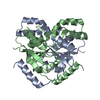
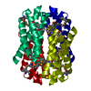
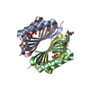
Mass: 18.015 Da / Num. of mol.: 137 / Source method: isolated from a natural source / Formula: H2O

-
Experimental details

-
Experiment

ExperimentMethod: X-RAY DIFFRACTION / Number of used crystals: 1

-
Sample preparation

CrystalDensity Matthews: 2.05 Å3/Da / Density % sol: 40 % / Description: NONE
Crystal growDetails: 200 MM NACL, 100 MM CITRATE PH 4, 800 MM AMMONIUM SULFATE

-
Data collection

DiffractionMean temperature: 100 K
Diffraction sourceSource: SYNCHROTRON / Site: Diamond / Beamline: I02 / Type: DIAMOND / Wavelength: 0.9796
DetectorType: DECTRIS PILATUS 6M / Detector: PIXEL / Date: Jan 26, 2014
RadiationProtocol: SINGLE WAVELENGTH / Monochromatic (M) / Laue (L): M / Scattering type: x-ray
Radiation wavelengthWavelength: 0.9796 Å / Relative weight: 1
ReflectionResolution: 1.7→45.41 Å / Num. obs: 64008 / % possible obs: 98.7 % / Observed criterion σ(I): 1.5 / Redundancy: 5.6 % / Rmerge(I) obs: 0.09 / Net I/σ(I): 8
Reflection shellResolution: 1.7→1.73 Å / Redundancy: 5.7 % / Rmerge(I) obs: 0.71 / Mean I/σ(I) obs: 1.5 / % possible all: 98

-
Processing

Software
NameVersionClassification
REFMAC5.8.0124refinement
XDSdata reduction
Aimlessdata scaling
MOLREPphasing
RefinementMethod to determine structure: MOLECULAR REPLACEMENT
Starting model: PDB ENTRY 5ARM
Resolution: 1.7→81.74 Å / Cor.coef. Fo:Fc: 0.936 / Cor.coef. Fo:Fc free: 0.914 / SU B: 6.128 / SU ML: 0.094 / Cross valid method: THROUGHOUT / ESU R: 0.124 / ESU R Free: 0.115 / Stereochemistry target values: MAXIMUM LIKELIHOOD / Details: HYDROGENS HAVE BEEN ADDED IN THE RIDING POSITIONS.
RfactorNum. reflection% reflectionSelection details
Rfree0.24262 3180 5 %RANDOM
Rwork0.21971 ---
obs0.22085 60813 99.7 %-
Solvent computationIon probe radii: 0.8 Å / Shrinkage radii: 0.8 Å / VDW probe radii: 1.2 Å / Solvent model: MASK
Displacement parametersBiso mean: 22.231 Å2
Baniso -1Baniso -2Baniso -3
1--0.27 Å20 Å20 Å2
2--0.59 Å20 Å2
3----0.32 Å2
Refinement stepCycle: LAST / Resolution: 1.7→81.74 Å
ProteinNucleic acidLigandSolventTotal
Num. atoms4320 0 0 137 4457
Refine LS restraints
Refine-IDTypeDev idealDev ideal targetNumber
X-RAY DIFFRACTIONr_bond_refined_d0.0160.0194466
X-RAY DIFFRACTIONr_bond_other_d0.0140.024101
X-RAY DIFFRACTIONr_angle_refined_deg1.8471.9536003
X-RAY DIFFRACTIONr_angle_other_deg2.3853.0029561
X-RAY DIFFRACTIONr_dihedral_angle_1_deg4.1645608
X-RAY DIFFRACTIONr_dihedral_angle_2_deg28.38825.249181
X-RAY DIFFRACTIONr_dihedral_angle_3_deg14.93215825
X-RAY DIFFRACTIONr_dihedral_angle_4_deg12.1321519
X-RAY DIFFRACTIONr_chiral_restr0.1020.2686
X-RAY DIFFRACTIONr_gen_planes_refined0.0110.025082
X-RAY DIFFRACTIONr_gen_planes_other0.0090.02921
X-RAY DIFFRACTIONr_nbd_refined
X-RAY DIFFRACTIONr_nbd_other
X-RAY DIFFRACTIONr_nbtor_refined
X-RAY DIFFRACTIONr_nbtor_other
X-RAY DIFFRACTIONr_xyhbond_nbd_refined
X-RAY DIFFRACTIONr_xyhbond_nbd_other
X-RAY DIFFRACTIONr_metal_ion_refined
X-RAY DIFFRACTIONr_metal_ion_other
X-RAY DIFFRACTIONr_symmetry_vdw_refined
X-RAY DIFFRACTIONr_symmetry_vdw_other
X-RAY DIFFRACTIONr_symmetry_hbond_refined
X-RAY DIFFRACTIONr_symmetry_hbond_other
X-RAY DIFFRACTIONr_symmetry_metal_ion_refined
X-RAY DIFFRACTIONr_symmetry_metal_ion_other
X-RAY DIFFRACTIONr_mcbond_it0.5320.7842372
X-RAY DIFFRACTIONr_mcbond_other0.530.7832371
X-RAY DIFFRACTIONr_mcangle_it0.911.1652949
X-RAY DIFFRACTIONr_mcangle_other
X-RAY DIFFRACTIONr_scbond_it1.0130.9452094
X-RAY DIFFRACTIONr_scbond_other
X-RAY DIFFRACTIONr_scangle_it
X-RAY DIFFRACTIONr_scangle_other
X-RAY DIFFRACTIONr_long_range_B_refined
X-RAY DIFFRACTIONr_long_range_B_other
X-RAY DIFFRACTIONr_rigid_bond_restr
X-RAY DIFFRACTIONr_sphericity_free
X-RAY DIFFRACTIONr_sphericity_bonded
LS refinement shellResolution: 1.7→1.744 Å / Total num. of bins used: 20
RfactorNum. reflection% reflection
Rfree0.28 225 -
Rwork0.279 4444 -
obs--99.34 %
Refinement TLS params.

Method: refined / Refine-ID: X-RAY DIFFRACTION

IDL112)L122)L132)L222)L232)L332)S11 (Å °)S12 (Å °)S13 (Å °)S21 (Å °)S22 (Å °)S23 (Å °)S31 (Å °)S32 (Å °)S33 (Å °)T112)T122)T132)T222)T232)T332)Origin x (Å)Origin y (Å)Origin z (Å)
12.4505-0.22020.33732.6907-0.15492.4436-0.078-0.1050.1079-0.07430.06610.1213-0.0042-0.22920.01190.0519-0.0763-0.01220.15310.00580.011716.72829.408945.7641
22.7964-0.4259-0.21462.57120.29932.6923-0.0944-0.0451-0.1779-0.09780.1226-0.0899-0.01480.2593-0.02820.0588-0.08520.01430.1569-0.00990.017337.725634.131145.0484
33.45810.17860.27172.4696-0.40292.90030.0508-0.1838-0.24460.1056-0.059-0.14270.26920.11740.00820.1549-0.0720.00490.13750.01780.031227.664820.258963.314
43.04130.4595-0.60732.66230.3783.31010.0386-0.16210.13560.0796-0.04030.1525-0.2444-0.15960.00180.1463-0.0682-0.00330.1292-0.0120.014928.738141.22964.0838
54.08010.02780.60731.74720.0053.0833-0.11650.1352-0.0926-0.11330.0971-0.109-0.11940.0560.01940.0973-0.06770.02380.0762-0.01430.01297.88948.272833.6669
63.6596-0.40560.52331.6276-0.15583.4973-0.1136-0.09740.120.08850.05310.0881-0.26230.06140.06050.09190.03340.00560.05320.00390.0121-7.16938.304351.8645
Refinement TLS group
IDRefine-IDRefine TLS-IDAuth asym-IDAuth seq-ID
1X-RAY DIFFRACTION1A4 - 108
2X-RAY DIFFRACTION2B2 - 108
3X-RAY DIFFRACTION3C4 - 108
4X-RAY DIFFRACTION4D4 - 108
5X-RAY DIFFRACTION5E4 - 108
6X-RAY DIFFRACTION6F4 - 108

+
About Yorodumi

-
News

-
Feb 9, 2022. New format data for meta-information of EMDB entries

New format data for meta-information of EMDB entries

  • Version 3 of the EMDB header file is now the official format.
  • The previous official version 1.9 will be removed from the archive.

Related info.:EMDB header

External links:wwPDB to switch to version 3 of the EMDB data model

-
Aug 12, 2020. Covid-19 info

Covid-19 info

URL: https://pdbj.org/emnavi/covid19.php

New page: Covid-19 featured information page in EM Navigator.

Related info.:Covid-19 info / Mar 5, 2020. Novel coronavirus structure data

+
Mar 5, 2020. Novel coronavirus structure data

Novel coronavirus structure data

Related info.:Yorodumi Speices / Aug 12, 2020. Covid-19 info

External links:COVID-19 featured content - PDBj / Molecule of the Month (242):Coronavirus Proteases

+
Jan 31, 2019. EMDB accession codes are about to change! (news from PDBe EMDB page)

EMDB accession codes are about to change! (news from PDBe EMDB page)

  • The allocation of 4 digits for EMDB accession codes will soon come to an end. Whilst these codes will remain in use, new EMDB accession codes will include an additional digit and will expand incrementally as the available range of codes is exhausted. The current 4-digit format prefixed with “EMD-” (i.e. EMD-XXXX) will advance to a 5-digit format (i.e. EMD-XXXXX), and so on. It is currently estimated that the 4-digit codes will be depleted around Spring 2019, at which point the 5-digit format will come into force.
  • The EM Navigator/Yorodumi systems omit the EMD- prefix.

Related info.:Q: What is EMD? / ID/Accession-code notation in Yorodumi/EM Navigator

External links:EMDB Accession Codes are Changing Soon! / Contact to PDBj

+
Jul 12, 2017. Major update of PDB

Major update of PDB

  • wwPDB released updated PDB data conforming to the new PDBx/mmCIF dictionary.
  • This is a major update changing the version number from 4 to 5, and with Remediation, in which all the entries are updated.
  • In this update, many items about electron microscopy experimental information are reorganized (e.g. em_software).
  • Now, EM Navigator and Yorodumi are based on the updated data.

External links:wwPDB Remediation / Enriched Model Files Conforming to OneDep Data Standards Now Available in the PDB FTP Archive

-
Yorodumi

Thousand views of thousand structures

  • Yorodumi is a browser for structure data from EMDB, PDB, SASBDB, etc.
  • This page is also the successor to EM Navigator detail page, and also detail information page/front-end page for Omokage search.
  • The word "yorodu" (or yorozu) is an old Japanese word meaning "ten thousand". "mi" (miru) is to see.

Related info.:EMDB / PDB / SASBDB / Comparison of 3 databanks / Yorodumi Search / Aug 31, 2016. New EM Navigator & Yorodumi / Yorodumi Papers / Jmol/JSmol / Function and homology information / Changes in new EM Navigator and Yorodumi

Read more