 Movie
Movie Controller


+ Open data

- Basic information
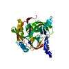
| Entry | Database: PDB / ID: 5fht | ||||||
|---|---|---|---|---|---|---|---|
| Title | HtrA2 protease mutant V226K | ||||||
 Components Components | Serine protease HTRA2, mitochondrial | ||||||
 Keywords Keywords |  HYDROLASE / APOPTOSIS HYDROLASE / APOPTOSIS | ||||||
| Function / homology |  Function and homology informationHtrA2 peptidase / pentacyclic triterpenoid metabolic process / negative regulation of mitophagy in response to mitochondrial depolarization / positive regulation of cysteine-type endopeptidase activity involved in apoptotic signaling pathway / regulation of autophagy of mitochondrion / ceramide metabolic process / CD40 receptor complex / : / programmed cell death / positive regulation of extrinsic apoptotic signaling pathway in absence of ligand ...HtrA2 peptidase / pentacyclic triterpenoid metabolic process / negative regulation of mitophagy in response to mitochondrial depolarization / positive regulation of cysteine-type endopeptidase activity involved in apoptotic signaling pathway / regulation of autophagy of mitochondrion / ceramide metabolic process / CD40 receptor complex / : / programmed cell death / positive regulation of extrinsic apoptotic signaling pathway in absence of ligand / serine-type endopeptidase complex / adult walking behavior / response to herbicide / positive regulation of protein targeting to mitochondrion / execution phase of apoptosis / positive regulation of cysteine-type endopeptidase activity involved in apoptotic process / protein autoprocessing / regulation of multicellular organism growth / negative regulation of cell cycle / neuron development / cellular response to interferon-beta / negative regulation of oxidative stress-induced intrinsic apoptotic signaling pathway / forebrain development / cellular response to retinoic acid / serine-type peptidase activity / mitochondrion organization / mitochondrial membrane / protein catabolic process / mitochondrial intermembrane space / cytoplasmic side of plasma membrane / cellular response to growth factor stimulus / intrinsic apoptotic signaling pathway in response to DNA damage / unfolded protein binding / cellular response to oxidative stress / cellular response to heat / peptidase activity / neuron apoptotic process / negative regulation of neuron apoptotic process / cytoskeleton / positive regulation of apoptotic process / serine-type endopeptidase activity / chromatin / endoplasmic reticulum membrane / endoplasmic reticulum / mitochondrion / proteolysis / membrane / identical protein binding / nucleus / cytosol Function and homology informationHtrA2 peptidase / pentacyclic triterpenoid metabolic process / negative regulation of mitophagy in response to mitochondrial depolarization / positive regulation of cysteine-type endopeptidase activity involved in apoptotic signaling pathway / regulation of autophagy of mitochondrion / ceramide metabolic process / CD40 receptor complex / : / programmed cell death / positive regulation of extrinsic apoptotic signaling pathway in absence of ligand ...HtrA2 peptidase / pentacyclic triterpenoid metabolic process / negative regulation of mitophagy in response to mitochondrial depolarization / positive regulation of cysteine-type endopeptidase activity involved in apoptotic signaling pathway / regulation of autophagy of mitochondrion / ceramide metabolic process / CD40 receptor complex / : / programmed cell death / positive regulation of extrinsic apoptotic signaling pathway in absence of ligand / serine-type endopeptidase complex / adult walking behavior / response to herbicide / positive regulation of protein targeting to mitochondrion / execution phase of apoptosis / positive regulation of cysteine-type endopeptidase activity involved in apoptotic process / protein autoprocessing / regulation of multicellular organism growth / negative regulation of cell cycle / neuron development / cellular response to interferon-beta / negative regulation of oxidative stress-induced intrinsic apoptotic signaling pathway / forebrain development / cellular response to retinoic acid / serine-type peptidase activity / mitochondrion organization / mitochondrial membrane / protein catabolic process / mitochondrial intermembrane space / cytoplasmic side of plasma membrane / cellular response to growth factor stimulus / intrinsic apoptotic signaling pathway in response to DNA damage / unfolded protein binding / cellular response to oxidative stress / cellular response to heat / peptidase activity / neuron apoptotic process / negative regulation of neuron apoptotic process / cytoskeleton / positive regulation of apoptotic process / serine-type endopeptidase activity / chromatin / endoplasmic reticulum membrane / endoplasmic reticulum / mitochondrion / proteolysis / membrane / identical protein binding / nucleus / cytosolSimilarity search - Function | ||||||
| Biological species |  Homo sapiens (human) Homo sapiens (human) | ||||||
| Method | X-RAY DIFFRACTION / MOLECULAR REPLACEMENT / Resolution: 1.95 Å | ||||||
 Authors Authors | Golik, P. / Dubin, G. / Zurawa-Janicka, D. / Lipinska, B. / Jarzab, M. / Wenta, T. / Gieldon, A. / Ciarkowski, A. | ||||||
 Citation Citation | Journal: Plos One / Year: 2016 Title: Distinct 3D Architecture and Dynamics of the Human HtrA2(Omi) Protease and Its Mutated Variants. Authors: Gieldon, A. / Zurawa-Janicka, D. / Jarzab, M. / Wenta, T. / Golik, P. / Dubin, G. / Lipinska, B. / Ciarkowski, J. | ||||||
| History |
|
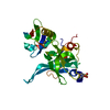
- Structure visualization
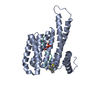
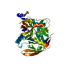
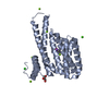
| Structure viewer | Molecule: MolmilJmol/JSmol |
|---|
- Downloads & links
-Download
| PDBx/mmCIF format | 5fht.cif.gz | 132.5 KB | Display | PDBx/mmCIF format |
|---|---|---|---|---|
| PDB format | pdb5fht.ent.gz | 102.3 KB | Display | PDB format |
| PDBx/mmJSON format | 5fht.json.gz | Tree view | PDBx/mmJSON format | |
| Others |  Other downloads Other downloads |
-Validation report
| Arichive directory | https://data.pdbj.org/pub/pdb/validation_reports/fh/5fhtftp://data.pdbj.org/pub/pdb/validation_reports/fh/5fht | HTTPS FTP |
|---|
-Related structure data
| Related structure data |  1lcyS S: Starting model for refinement |
|---|---|
| Similar structure data |


-Links
 PDBj
PDBj- Assembly
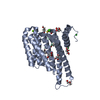
| Deposited unit | 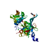
| ||||||||
|---|---|---|---|---|---|---|---|---|---|
| 1 |
| ||||||||
| Unit cell |
| ||||||||
| Components on special symmetry positions |
|
-Components
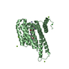
-Protein , 1 types, 1 molecules A
| #1: Protein | Mass: 36234.207 Da / Num. of mol.: 1 Source method: isolated from a genetically manipulated source Source: (gene. exp.) Homo sapiens (human) / Gene: HTRA2, OMI, PRSS25 / Production host:  Escherichia coli BL21(DE3) (bacteria) / References: UniProt: O43464, HtrA2 peptidase Escherichia coli BL21(DE3) (bacteria) / References: UniProt: O43464, HtrA2 peptidase |
|---|
-Non-polymers , 5 types, 204 molecules 


| #2: Chemical | ChemComp-MES / MES (buffer) Mass: 195.237 Da / Num. of mol.: 1 / Source method: obtained synthetically / Formula: C6H13NO4S / Comment: pH buffer*YM Mass: 195.237 Da / Num. of mol.: 1 / Source method: obtained synthetically / Formula: C6H13NO4S / Comment: pH buffer*YM |
|---|---|
| #3: Chemical | ChemComp-CL / Chloride Mass: 35.453 Da / Num. of mol.: 1 / Source method: obtained synthetically / Formula: Cl Mass: 35.453 Da / Num. of mol.: 1 / Source method: obtained synthetically / Formula: Cl |
| #4: Chemical | ChemComp-K /  Mass: 39.098 Da / Num. of mol.: 1 / Source method: obtained synthetically / Formula: K Mass: 39.098 Da / Num. of mol.: 1 / Source method: obtained synthetically / Formula: K |
| #5: Chemical | ChemComp-NA /  Mass: 22.990 Da / Num. of mol.: 1 / Source method: obtained synthetically / Formula: Na Mass: 22.990 Da / Num. of mol.: 1 / Source method: obtained synthetically / Formula: Na |
| #6: Water | ChemComp-HOH / WaterMass: 18.015 Da / Num. of mol.: 200 / Source method: isolated from a natural source / Formula: H2O |
-Experimental details
-Experiment
| Experiment | Method: X-RAY DIFFRACTION |
|---|
- Sample preparation
| Crystal | Density Matthews: 2.49 Å3/Da / Density % sol: 50.58 % |
|---|---|
| Crystal grow | Temperature: 293 K / Method: vapor diffusion, sitting drop / Details: MES, NaCl, KH2PO4, NaH2PO4 / PH range: 6.5 |
-Data collection
| Diffraction | Mean temperature: 77 K |
|---|---|
| Diffraction source | Source: ROTATING ANODE / Type: RIGAKU MICROMAX-007 HF / Wavelength: 1.54 Å |
| Detector | Type: RIGAKU RAXIS IV++ / Detector: IMAGE PLATE / Date: Dec 17, 2012 |
| Radiation | Protocol: SINGLE WAVELENGTH / Monochromatic (M) / Laue (L): M / Scattering type: x-ray |
| Radiation wavelength | Wavelength: 1.54 Å / Relative weight: 1 |
| Reflection | Resolution: 1.95→14.57 Å / Num. obs: 25396 / % possible obs: 99.6 % / Redundancy: 4.8 % / Rmerge(I) obs: 0.116 / Net I/σ(I): 7.4 |
| Reflection shell | Highest resolution: 1.95 Å / Redundancy: 4.3 % / Rmerge(I) obs: 0.405 / Mean I/σ(I) obs: 3.1 / % possible all: 99.5 |
- Processing
| Software |
| ||||||||||||||||||||||||||||||||||||||||||||||||||||||||||||||||||||||||||||||||||||||||||||||||||||||||||||||||||||||||||||||||||||||||||||||||||||||||||||||||||||||||||||||||||||||
|---|---|---|---|---|---|---|---|---|---|---|---|---|---|---|---|---|---|---|---|---|---|---|---|---|---|---|---|---|---|---|---|---|---|---|---|---|---|---|---|---|---|---|---|---|---|---|---|---|---|---|---|---|---|---|---|---|---|---|---|---|---|---|---|---|---|---|---|---|---|---|---|---|---|---|---|---|---|---|---|---|---|---|---|---|---|---|---|---|---|---|---|---|---|---|---|---|---|---|---|---|---|---|---|---|---|---|---|---|---|---|---|---|---|---|---|---|---|---|---|---|---|---|---|---|---|---|---|---|---|---|---|---|---|---|---|---|---|---|---|---|---|---|---|---|---|---|---|---|---|---|---|---|---|---|---|---|---|---|---|---|---|---|---|---|---|---|---|---|---|---|---|---|---|---|---|---|---|---|---|---|---|---|---|
| Refinement | Method to determine structure: MOLECULAR REPLACEMENT Starting model: poly-alanine model of 1LCY Resolution: 1.95→14.57 Å / Cor.coef. Fo:Fc: 0.955 / Cor.coef. Fo:Fc free: 0.929 / SU B: 5.865 / SU ML: 0.086 / Cross valid method: THROUGHOUT / ESU R: 0.146 / ESU R Free: 0.146 / Stereochemistry target values: MAXIMUM LIKELIHOOD / Details: HYDROGENS HAVE BEEN ADDED IN THE RIDING POSITIONS
| ||||||||||||||||||||||||||||||||||||||||||||||||||||||||||||||||||||||||||||||||||||||||||||||||||||||||||||||||||||||||||||||||||||||||||||||||||||||||||||||||||||||||||||||||||||||
| Solvent computation | Ion probe radii: 0.8 Å / Shrinkage radii: 0.8 Å / VDW probe radii: 1.2 Å / Solvent model: MASK | ||||||||||||||||||||||||||||||||||||||||||||||||||||||||||||||||||||||||||||||||||||||||||||||||||||||||||||||||||||||||||||||||||||||||||||||||||||||||||||||||||||||||||||||||||||||
| Displacement parameters | Biso mean: 34.372 Å2
| ||||||||||||||||||||||||||||||||||||||||||||||||||||||||||||||||||||||||||||||||||||||||||||||||||||||||||||||||||||||||||||||||||||||||||||||||||||||||||||||||||||||||||||||||||||||
| Refinement step | Cycle: LAST / Resolution: 1.95→14.57 Å
| ||||||||||||||||||||||||||||||||||||||||||||||||||||||||||||||||||||||||||||||||||||||||||||||||||||||||||||||||||||||||||||||||||||||||||||||||||||||||||||||||||||||||||||||||||||||
| Refine LS restraints |
|